No products in the cart.
RNASweAMI™ In Situ Hybridization Detection Kits
Description
Call or send Email for discount ! sure your satisfy!
| Product Code | Product Name | Size | |
|---|---|---|---|
| GF001-50T | RNASweAMI™ In Situ Hybridization DAB Detection Kit | 50 T | $3000 |
| GF002-50T | RNASweAMI™ In Situ Hybridization CY3 Detection Kit | 50 T | $3000 |
| GF003-50T | RNASweAMI™ In Situ Hybridization IF488 Detection Kit | 50 T | $3000 |
| GF004-50T | RNASweAMI™ In Situ Hybridization IF550 Detection Kit | 50 T | $3000 |
| GF005-50T | RNASweAMI™ In Situ Hybridization IF590 Detection Kit | 50 T | $3000 |
| GF006-50T | RNASweAMI™ In Situ Hybridization IF647 Detection Kit | 50 T | $3000 |
| GF007-50T | RNASweAMI™ In Situ Hybridization Multi-fluorescence Detection Kit (IF488-550) | 50 T | $4500 |
| GF008-50T | RNASweAMI™ In Situ Hybridization Multi-fluorescence Detection Kit (IF488-550-647) | 50 T | $6000 |
| GF009-50T | RNASweAMI™ In Situ Hybridization DAB Signal Detection Kit (Probe not included) | 50 T | $1500 |
| GF010-50T | RNASweAMI™ In Situ Hybridization Fluorescence Signal Detection Kit (Probe not included) | 50 T | $1500 |
| G3045-30ML | Hybridization Buffer (10% formamide) | 30 mL | $100 |
| G3046-30ML | Hybridization Buffer (20% formamide) | 30 mL | $100 |
| G3047-30ML | Hybridization Buffer (30% formamide) | 30 mL | $100 |
| G3048-30ML | Hybridization Buffer (40% formamide) | 30 mL | $100 |
| G3049-30ML | Hybridization Buffer (50% formamide) | 30 mL | $100 |
| G3051-30ML | Hybridization Buffer (15% formamide) | 30 mL | $100 |
| G3052-100ML | 20×SSPE Buffer | 100 mL | $50 |
| G3053-100ML | 50×Denhardt’s solution | 100 mL | $120 |
| GP00006 | RNASweAMI Human Positive Control Probe (Hs-RNU6-1) | 50 μL | $100 |
| GP00007 | RNASweAMI Mouse Positive Control Probe (Mm-Rnu6) | 50 μL | $100 |
1.
GF001-50T RNASweAMI™ In Situ Hybridization DAB Detection Kit – Composition and Storage Conditions
| Component No. | Component Name | 50T Size | Storage |
|---|---|---|---|
| GP-####-1a | Target Probe-[species]-[gene]-Mix1 | 650 μL | -20℃ |
| GP-####-2a | Target Probe-[species]-[gene]-Mix2 | 650 μL | -20℃ |
| G3701-25UL | DIG probe (200×) | 25 μL | -20℃ |
| G3702-15UL | anti-DIG (HRP) | 15 μL | -20℃ |
| G3708-100ML | Fixation Solution | 100 mL | 4℃ |
| G3709-50ML | Fixation Solution (20×) | 50 mL | 4℃ |
| G3710-1ML | Proteinase K | 1 mL | -20℃ |
| G3711-20ML | Permeabilization Reagent b | 20 mL | 4℃ |
| G3712-50ML | Hybridization Buffer | 50 mL | 4℃ |
| G3713-100ML | 20×SSC | 300 mL | 4℃ |
| G3716-10ML | Blocking Solution | 10 mL | 4℃ |
| G3717-100UL | DAB Color Development Reagent A | 100 μL | 4℃ |
| G3718-10ML | DAB Color Development Reagent B | 10 mL | 4℃ |
Note:
- a The ##### in the target probe’s product code represents a unique numeric code for each probe. Species abbreviations: Mm=mouse, Hs=human, Rn=rat, Oc=rabbit, Bt=bovine, Dr=zebrafish.
- b The permeabilization reagent contains 0.5% Triton X-100. The concentration can be adjusted based on the cell type. If a lower concentration of Triton X-100 is required, it can be diluted with RNase-free 1×PBS without nucleases.
Sizec: The entire kit can be used to detect 50 tissue spots with an approximate area of 20 mm×20 mm each. When used for larger sample areas, the number of experiments per kit will decrease accordingly.
Storage: All reagents have a shelf life of 12 months under storage conditions. The entire kit is transported in a low-temperature ice bag.
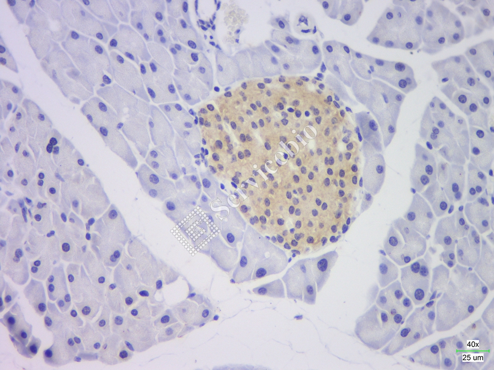
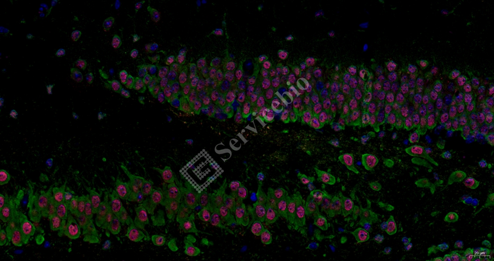
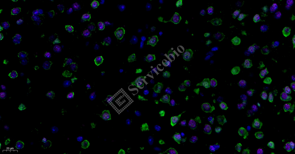
Introduction to RNASweAMI In Situ Hybridization: In situ hybridization (ISH) is based on the principle of base complementarity pairing. It utilizes specific labeled single-stranded nucleic acids (probes) with known sequences, such as fluorescein or digoxigenin, to specifically bind to the target gene (DNA or RNA) in cells or tissue sections. The hybridization of the target gene and the labeled probe forms a hybrid body. Subsequently, depending on the label of the probe, different color development systems are used to visualize and amplify the signal of the hybrid body, enabling precise localization of the target gene. Depending on the probe label, there are fluorescence color development systems, DAB color development systems, etc.
RNASweAMI ISH In Situ Hybridization Detection Procedure: Please refer to the provided figure for an overview of the experimental procedure.
User-supplied materials (as shown in Table 1 in the provided document): This section provides a list of materials that users need to prepare for the RNASweAMI ISH experiment, including reagents, solvents, and equipment. The user needs to obtain or prepare these materials separately as they are not included in the kit.
Please note that this product is for research use only and not intended for clinical diagnosis.
Please let me know if you need any further information or clarification.
2. GF002-50TRNASweAMI™ In Situ Hybridization CY3 Detection Kit
2.
| Component No. | Component name | 50T Size | Storage |
|---|---|---|---|
| GP-####-1a | Target Probe-[species]-[gene]-Mix1 | 650 μL | -20°C |
| GP-####-2a | Target Probe-[species]-[gene]-Mix2 | 650 μL | -20°C |
| G3703-25UL | CY3 probe (200×) | 25 μL | -20°C |
| G3708-100ML | Fixing Solution | 100 mL | 4°C |
| G3709-50ML | Fixation Solution (20×) | 50 mL | 4°C |
| G3710-1ML | Proteinase K | 1 mL | -20°C |
| G3711-20ML | Permeabilization Reagent b | 20 mL | 4°C |
| G3712-50ML | Hybridization Solution | 50 mL | 4°C |
| G3713-100ML | 20×SSC | 300 mL | 4°C |
| G3714-25ML | DAPI solution | 25 mL | 4°C |
| G3715-25ML | Anti-fluorescence quenching sealing agent | 25 mL | -20°C |
a The #### in the target probe catalog number represents a unique numerical code for each probe, and the species abbreviations are: Mm = mouse, Hs = human, Rn = rat, Oc = Rabbit, Bt = Bovine, Dr = zebrafish.
b The effective component of the permeabilization reagent is 0.5% Triton X-100. The concentration can be adjusted for different cells. To achieve a lower concentration of Triton X-100 for cell permeabilization, dilute the reagent with RNase-free 1×PBS.
c The entire set of reagents can be used to test 50 tissue spots with an area of approximately 20 mm×20 mm each. If used for larger sample areas, the number of experiments that can be completed per set of reagents will be reduced accordingly.
d All reagents have a shelf life of 12 months under storage conditions. The entire kit is transported in a low-temperature ice bag.
Introduction to RNASweAMI in situ hybridization (ISH)
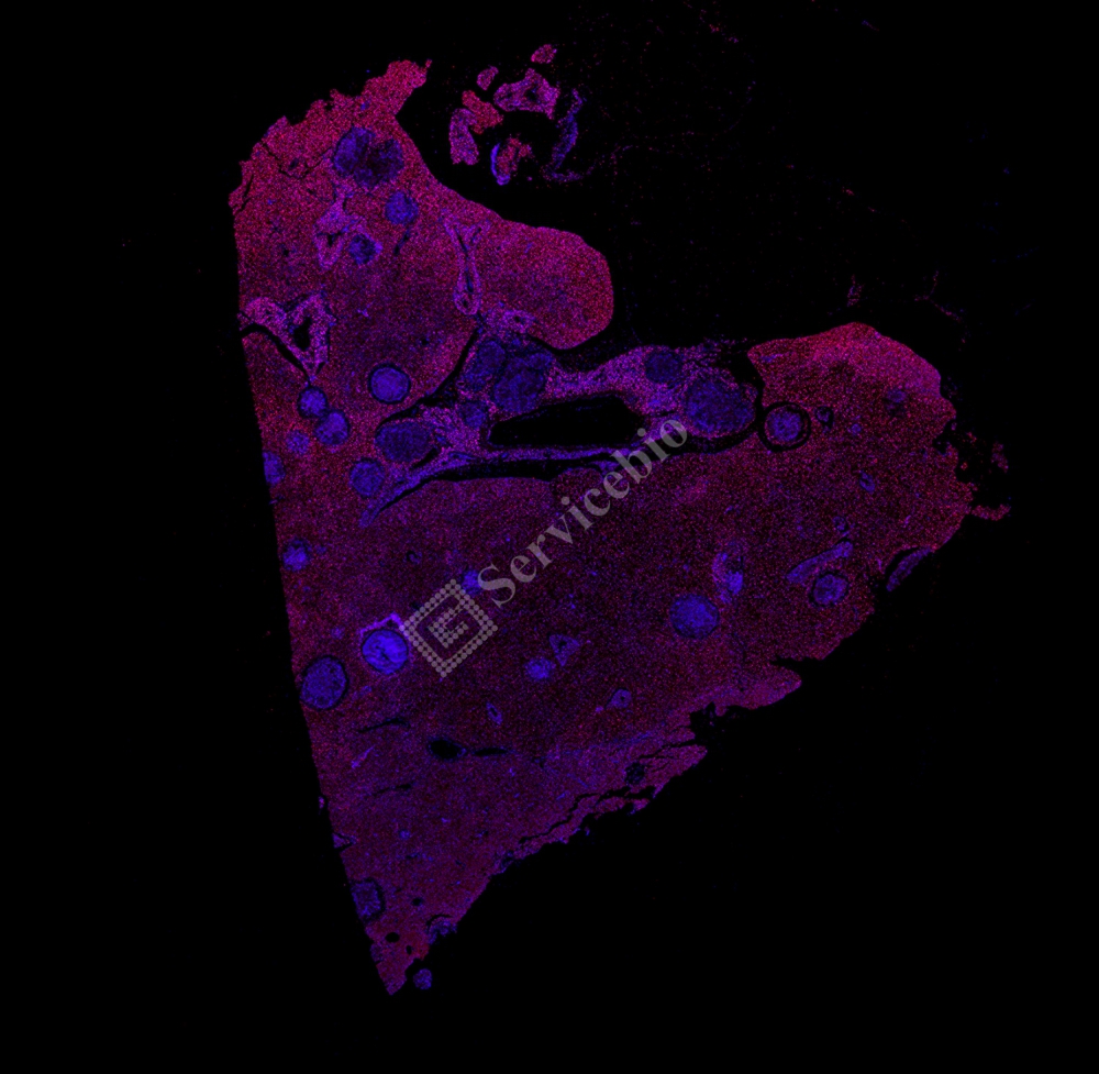
In situ hybridization (ISH) is a technique based on the principle of base complementarity. It uses specific labeled single-stranded nucleic acids (probes), such as fluorescent or digoxigenin-labeled sequences, to bind specifically to the target genes (DNA or RNA) in cells or tissue sections to form a hybrid complex through base complementarity. Then, based on the label of the probe, various colorimetric or fluorescence-based systems are used to visualize and amplify the hybridization signal, allowing for precise localization of the target genes. Depending on the label of the probe, there are fluorescence colorimetric systems, DAB colorimetric systems, and others.
RNASweAMI ISH in situ hybridization technology follows the detection process illustrated in Figure 1. Specific oligonucleotide probes bind to the target RNA in the tissue sample through base complementarity and are subsequently enriched by two “L-shaped” structures (single-stranded DNA), achieving a cascade signal amplification. Afterward, bright-field or fluorescence imaging techniques can be used to observe the apparent dot-like or clustered signals formed at the target RNA site. Compared to traditional ISH labeling methods, this method theoretically allows signal amplification up to 400-8000 times, enabling the visualization of single-molecule RNA with a high signal-to-noise ratio.
3.