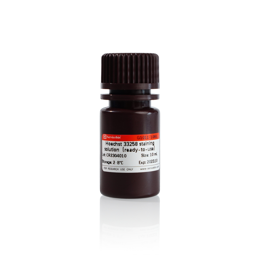
Description
Product Information
| Product Name | Cat.No. | Spec. |
| JC-1 Mitochondrial Membrane Potential Assay Kit | G1515-100T | 100T |
Description
JC-1 is a cationic carbonyl cyanine dye, which can pass through the cell membrane and aggregate towards mitochondria under the action of mitochondrial membrane potential. It is an ideal fluorescent probe widely used in detecting mitochondrial membrane potential.When the mitochondrial membrane potential is high, the dye aggregates in the matrix of mitochondria,showing red fluorescence(Ex=585 nm,Em=590 nm);When the mitochondrial membrane potential is low, JC-1 is predominantly a monomer exist in cytoplasm,and showing green fluorescence(Ex=514 nm,Em=529 nm).Therefore, the change of mitochondrial membrane potential can be judged according to the transformation of fluorescence color and the change of proportion, and the decrease of mitochondrial membrane potential is also an important marker of early apoptosis.
JC-1 Mitochondrial Membrane Potential Assay Kit,is based on JC-1 fluorescent probe,which has improved easy precipitation problem and other shortcomings.This kit can be used to quickly and sensitively detect mitochondrial membrane potential changes in cell, tissue or purified mitochondria, and is also a common method used to detect early cell apoptosis.
In addition, this kit also provides CCCP (Carbonyl cyanide 3-chlorophenylhydrazone) reagent, a proton carrier (hydrogen ionophore) and decoupler of oxidative phosphorylation in mitochondria, which can promote changes in the permeability of mitochondria to hydrogen ions , resulting in the decrease or loss of mitochondrial membrane potential, which can be used as a positive control for inducing the decrease of mitochondrial membrane potential.
Storage and Handling Conditions
JC-1 dye(500×)should be stored at -20℃,desiccated and protected from light,and avoid repeated freezing and thawing;
JC-1 buffer and JC-1 diluent may be stored at 4℃.
Component
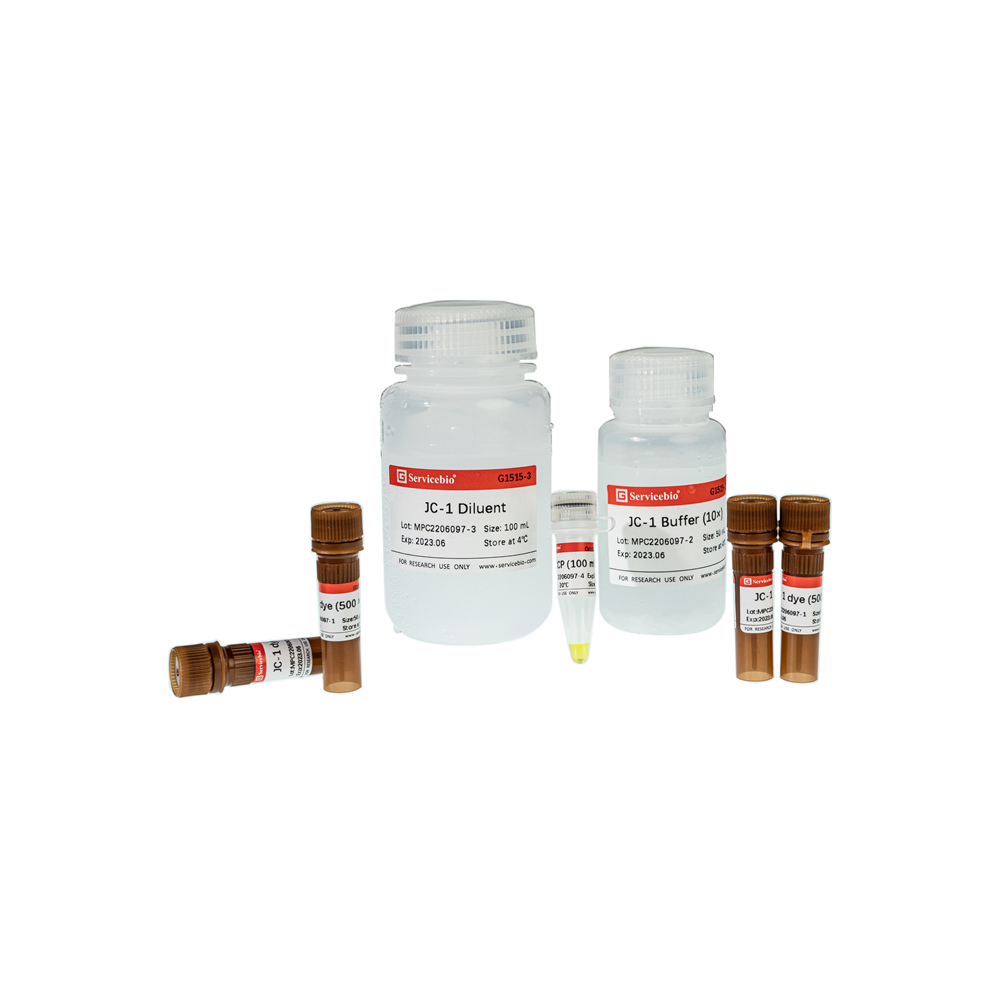
| Component Number | Component | G1515-100T |
| G1515-1 | JC-1 dye(500 ×) | 4 x 50 μL |
| G1515-2 | JC-1 buffer (10 x) | 50 mL |
| G1515-3 | JC-1 diluent | 100 mL |
| G1515-4 | CCCP(100 mM) | 50 μL |
Assay Protocol / Procedures
1. prepare the working solution of JC-1 dye(2x) and JC-1 buffer(1x):
a. In a suitable container mix 2 μL JC-1 dye(500 ×)to 900 μL JC-1 diluent,then add 100 μL JC-1 buffer (10 x).(Note:this solution should be prepared in order.)
b. In a suitable container diluent the JC-1 buffer (10 x) with ddH2O.
2. prepare a positive control(optional ):
a. Take an appropriate amount of CCCP (100 mM) and dilute 1000 times with cell culture medium to obtain 100 μM CCCP working solution;
b. Incubate the cells with CCCP working solution for about 30 minutes, and then follow the JC-1 staining procedure below to detect the mitochondrial membrane potential.
Note: For different cells, the concentration and incubation time of CCCP may be different, please refer to reference or do experiment to find the optimal conditions.
3. Cell stain and analyze
a. Take 100,000 to 600,000 cells, wash cells in JC-1 buffer(1x) or PBS;
b. Add JC-1 dye(2x) and cell culture medium with a ratio of 1:1 to the cells,and incubate in a CO2 incubator for 15-30 min in the dark;
c. Wash cells twice with 1x JC-1 buffer;
d. Analyze on a fluorescence microscope, laser confocal microscope, or flow cytometry.If the cell mainly shows red fluorescence, it indicates that its mitochondrial membrane potential is normal; if the green fluorescence is greatly increased, it indicates that the mitochondrial membrane potential has decreased, and it may be in the early stage of cell apoptosis.
4. Mitochondria stain and analyze
a. Add 100 μL of JC-1 staining working solution to 100 μL of purified mitochondria with a total protein content of 10-100 μg;
b. After mixing, use a fluorescence spectrophotometer (excitation wavelength is 485 nm, emission wavelength is 590 nm) or microplate reader and other instruments for scanning detection (when the excitation wavelength cannot be set to 485 nm, it can be set in the range of 475 ~ 520 nm Excitation wavelength), can also be observed by fluorescence microscope or laser confocal microscope
Note: For the accuracy of the experiment, it is recommended that the samples after JC-1 incubation should be tested within 30 minutes.
Here is translate from Chinese page, it may help your idea.
Operation Steps:
- Preparation of JC-1 Staining Working Solution and JC-1 Staining Buffer:
a. Take 2 μL of JC-1 solution (500×) and mix thoroughly with 900 μL of JC-1 dilution solution. Then add 100 μL of JC-1 staining buffer (10×) and vortex to mix well. This will prepare the JC-1 staining working solution (note the order of adding the components).
b. Take an appropriate amount of JC-1 staining buffer (10×) and dilute it with deionized water to prepare a 1× JC-1 staining buffer (unless otherwise specified, use 1× JC-1 staining buffer for washing steps).
- Positive Control Group Setup:
a. Take an appropriate amount of CCCP (100 mM) and dilute it 1000 times with cell culture medium to obtain a 100 μM CCCP working solution.
b. Incubate the cells with the CCCP working solution for about 30 minutes, then proceed to the JC-1 staining steps below to measure the mitochondrial membrane potential.
(Note: For most cells, after the above treatment, compared to the normal group, CCCP treatment will result in JC-1 mostly existing as monomers in the cytoplasm, showing bright green fluorescence and reduced red fluorescence. However, for some cells, the concentration and duration of CCCP treatment may vary. Please refer to relevant literature or optimize the system to determine the optimal conditions.)
- Staining of Suspended Cells:
a. Take 1-6×10^5 cells, centrifuge at 1000 g for 3-5 minutes to remove the original culture medium, add JC-1 staining buffer for one wash, then centrifuge to remove the JC-1 staining buffer, and resuspend in 500 μL of cell culture medium (can contain serum and phenol red).
b. Then add 500 μL of JC-1 staining working solution, protect from light, and incubate in a CO2 incubator for 15-30 minutes (generally, 20 minutes is sufficient, but the incubation time can be adjusted as needed).
c. After incubation, proceed with the usual procedure for suspended cells, centrifuge to remove the JC-1 staining working solution, and wash twice with JC-1 staining buffer.
d. Resuspend in an appropriate amount of JC-1 staining buffer (1×) or cell culture medium (recommended without phenol red), and observe under a fluorescence microscope or confocal laser scanning microscope. Alternatively, use flow cytometry or other instruments for analysis.
- Staining of Adherent Cells (Using a 6-well plate as an example):
For adherent cells, if flow cytometry analysis is required, collect the cells and follow the detection method for suspended cells.
a. Remove the original medium containing drugs or other treatments, add 1 mL of JC-1 staining buffer, and wash 1-2 times.
b. Add 1 mL of cell culture medium (can contain serum and phenol red).
c. Add 1 mL of JC-1 staining working solution, gently mix, protect from light, and incubate in a CO2 incubator for 15-30 minutes.
d. After incubation, remove the supernatant, wash twice with JC-1 staining buffer.
e. Add 2 mL of JC-1 staining buffer (1×) or cell culture medium (recommended without phenol red).
f. Observe under a fluorescence microscope or confocal laser scanning microscope, or use flow cytometry or other instruments for analysis.
- Measurement of Purified Mitochondria:
a. Add 100 μL of purified mitochondria (witha total protein content of 10-100 μg) to 900 μL of JC-1 staining working solution.
b. Mix well and perform scanning detection using a fluorescence spectrophotometer (excitation wavelength: 485 nm, emission wavelength: 590 nm) or an ELISA reader (if the excitation wavelength cannot be set to 485 nm, set it within the range of 475-520 nm). Alternatively, observe using a fluorescence microscope or confocal laser scanning microscope. Refer to the wavelength settings in Step 6 for detection.
- Fluorescence Detection and Analysis:
a. The wavelength parameters for detecting JC-1 monomers are Ex=490 nm, Em=525 nm, and for JC-1 aggregates are Ex=525 nm, Em=590 nm. Note that the excitation and emission wavelengths for fluorescence detection do not have to be set at the maximum excitation and emission wavelengths. Adjust the parameters accordingly based on the specific situation.
b. When taking fluorescence microscope images, it is recommended to use the normal group as a standard to determine the exposure time for red and green fluorescence. The red fluorescence of the normal group should be clear, while the green fluorescence should be relatively dim. Take images of the fluorescence in the treated groups using the same exposure time as the normal group.
c. Interpretation of results: In normal cell conditions, predominantly red fluorescence indicates normal mitochondrial membrane potential. If there is a significant increase in green fluorescence, it indicates a decrease in mitochondrial membrane potential and the cells may be in the early stage of apoptosis.
Notes:
- To ensure complete dissolution of JC-1, follow the specified order of operations for preparing the JC-1 working solution.
- It is recommended to complete the detection of samples incubated with JC-1 within 30 minutes.
- If there is precipitation in the JC-1 solution or JC-1 staining buffer (10×), gently warm and dissolve it in a water bath if necessary.
- For the final JC-1 detection, it is recommended to use cell culture medium without phenol red to avoid interference from background color. Phenol red has no effect on the results during the JC-1 staining and incubation stages.
- If the amount of JC-1 solution used each time is small and multiple freeze-thaw cycles are involved, consider aliquoting the solution to minimize repeated freeze-thaw cycles of the JC-1 solution.
- Wear laboratory attire and disposable gloves during the operation.
This product is intended for research use only and is not for clinical diagnosis.
For Research Use Only!
|
Cat.No.
|
Product Name
|
Spec.
|
Operation
|
|---|
|
G1011-10ML
|
Hoechst 33258 Staining Solution (Ready-To-Use)
|
10 mL
|
|
|
G1012-100ML
|
DAPI Staining Reagent (Ready to Use)
|
100 mL
|
|
|
G1012-10ML
|
DAPI Staining Reagent (Ready to Use)
|
10 mL
|
|
|
G1021-10ML
|
PI Stain
|
10 mL
|
|
|
G1127-1ML
|
Hoechst 33342 Staining Solution
|
1 mL
|
|
|
G1515-100T
|
JC-1 Mitochondrial Membrane Potential Assay Kit
|
100 T
|
|
|
G1704
|
DiO Cell Membrane Green Fluorescence Staining Kit
|
100-1000 T
|
|
|
G1705
|
Dil Cell Membrane Red Fluorescence Staining Kit
|
100-1000 T
|

