No products in the cart.


5. TSAPLus Fluorescence Triple Staining Kit (C type), (Plus Fluorescence Triple Staining), 100T 50μL , $1800
$2,500.00
In Stock
Description
Product Information
| Product Name | Cat.No. | Spec. |
| TSAPLus Fluorescence Triple Staining Kit | G1236-50T | 50T |
| G1236-100T | 100T |
Description
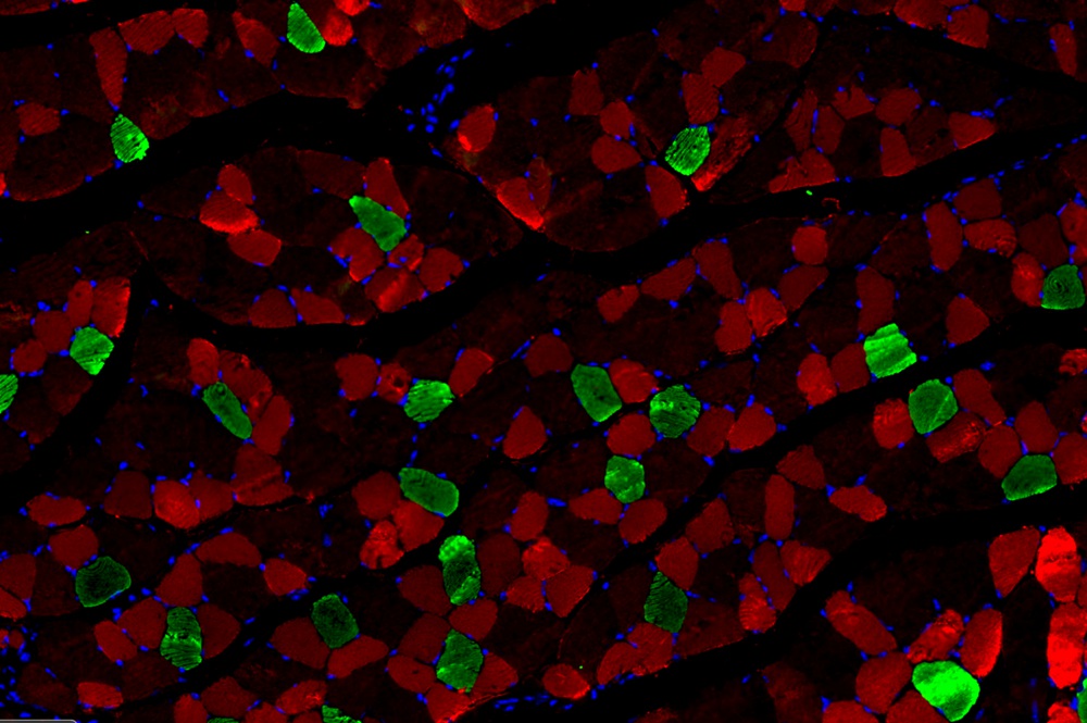
This TSAPLus Fluorescence Triple Staining Kit is used for multiple immunofluorescence staining of paraffin sections, especially for multiple immunofluorescence labeling of primary antibodies from the same source. The principle of this kit is based on the tyramide signal amplification technology, short for TSA. In the TSA technology, during the catalytic activity of HRP (horseradish peroxidase), fluorescence labeled tyramine was also catalyzed. The resulting enzymatic reaction products bind to surrounding protein residues (including tryptophan, histidine and tyrosine residues) in situ, which form a large amount of fluorescein deposition at the antigen-antibody binding site to achieve signal amplification. This kit uses fluorescent dye 488/555/647 to label tyramine, and the obtained fluorescent tyramine has strong fluorescence and stable signal, which can be used for multiple repeated immunolabeling to achieve multiple fluorescence staining.
Fluorescence spectrum data of fluorescent dyes associated with TSA series fluorescence staining kits are as follows:
| Fluorescence Labeled Tyramine | Ex/Em |
| FITC-Tyramide | 492/518 |
| CY3-Tyramide | 555/569 |
| iF488-Tyramide | 491/516 |
| iF555-Tyramide | 557/570 |
| iF647-Tyramide | 656/670 |
Storage and Handling Conditions
Each component in the kit is stored according to the required conditions; the whole kit is transported in wet ice. Valid for one year.
Component
| Component Number | Component | G1236-50T | G1236-100T | Storage |
| G1236-1 | iF488-Tyramide | 25 μL | 50 μL | -20℃ |
| G1236-2 | iF555-Tyramide | 25 μL | 50 μL | -20℃ |
| G1236-3 | iF647-Tyramide | 25 μL | 50 μL | -20℃ |
| G1236-4 | Tyramide Dilution Buffer | 100 mL | 2×100 mL | 4℃ |
| G1236-5 | DAPI(ready-to-use) | 10 mL | 20 mL | 4℃ |
| G1236-6 | Tissue Autofluorescence Quencher (Solution A) | 10 mL | 20 mL | RT |
| G1236-7 | Tissue Autofluorescence Quencher (Solution B) | 10 mL | 20 mL | RT |
| G1236-8 | Anti fluorescence Quenching Biomount | 5 mL | 10 mL | -20℃ |
| Manual | One cppy | |||
Experiment Preparation
1. Prepare 0.01 mol/L PBS buffer (pH7.0-7.4, recommended G4202 or G0002), 3% H2O2 and 0.3% H2O2;
2. Prepare primary antibody and corresponding HRP labeled secondary antibody, antigen repair solution (select appropriate antigen repair solution according to antibody and tissue type)
3. According to the dosage, prepare TSA working solution according to the following proportion.
| TSA Working Solution | Reagent Name | Volume |
| TSA-488 working solution | Tyramide Dilution Buffer | 1 mL |
| 0.3% H2O2 | 10 μL | |
| iF488-Tyramide | 2 μL | |
| TSA-555 working solution | Tyramide Dilution Buffer | 1 mL |
| 0.3% H2O2 | 10 μL | |
| iF555-Tyramide | 2 μL | |
| TSA-647 working solution | Tyramide Dilution Buffer | 1 mL |
| 0.3%H2O2 | 10 μL | |
| iF647-Tyramide | 2 μL |
Note: centrifuge the fluorescence tyramide before use; the TSA working solution is recommended to be prepared on the spot, and should be stored at 4℃ protected from light, and this solution is effective within 24 h.
Usage
1. Prepare Sample: deparaffinize the tissue paraffin section and dehydrate according to standard IHC protocols.
2. (Optional) Antigen repair: According to the sample type and primary antibody used, antigen repair was performed on the tissue sections in an appropriate manner. Then rinse the tissue three times in PBS 1× at room temperature.
3. Draw a circle around tissue use a hydrophobic barrier pen to hold liquid reagents.
4. (Optional) If the spontaneous fluorescence is needed to quench , add 100 μL Tissue Autofluorescence Quencher (Solution A) to cover the sample and incubate for 30 min at room temperature. Then rinse the sample with water for 5 min.
5. For quench the endogenous peroxidase activity, add enough drops of 3% H2O2 to cover the sample and incubate protect from light for 25 min at room temperature.
6. Blocking: add 100-150 μL of 3% BSA or serum to cover the sample and incubate for 30 min. Select blocking reagent according to primary and secondary antibody species.
7. The first fluorescent label 488
7.1 Label the tissue with the first primary antibody: Gently shake off the liquid on the section, drop the diluted primary antibody (Dilute primary antibody to appropriate concentration with PBS ) to cover the tissue, and place the section flat in a wet box with water and incubate at 4℃ overnight.
7.2 Rinse the cells or tissue for 5 min with PBS 1× at room temperature. Repeat this step three times.
7.3 The first secondary antibody reaction: add 100-150 μL of the primary antibody corresponded HRP-conjugated secondary antibody to the cells or tissue for 50 min at room temperature.
Note: if you observe non-specific signal, you can short this incubation time.
7.4 Rinse the cells or tissue for 5-10 min with PBS 1× at room temperature. Repeat this step three times.
7.5 TSA fluorescence label: apply 100 μL of the TSA-488 working solution to the tissue and incubate for 10 min protect from light. Rinse the tissue three times with PBS 1×, 5 min each.
8. Heat treatment: Tissue sections were placed in a repair box filled with antigenic repair solution (appropriate antigenic repair solution was selected according to tissue type and antibody) for microwave heating to remove the primary and secondary antibodies that had been bound. In this step, dry sheets caused by excessive evaporation of liquid should be prevented. At this step, the first fluorescent label 488 is completed.
9. The second fluorescent label 555: Repeat the step 7 and step 8 to label the tissue with the second primary antibody and corresponded HRP-conjugated secondary antibody, TSA-555 working solution, and then heat treatment to remove primary-secondary antibody complex. At this step, the first fluorescent label 555 is completed.
10. The second fluorescent label 647: In the same way, repeat the step 7 to label the tissue with the third primary antibody and corresponded HRP-conjugated secondary antibody, TSA-647 working solution, and then heat treatment to remove primary-secondary antibody complex. At this step, the first fluorescent label 647 is completed.
11. Nuclear counterstain: add 100-150 μL DAPI (ready-to-use) to cover the tissue and incubate at room temperature protect from light for 10 min.
12. Rinse the tissue three times with PBS 1×, 5 min each.
13. (Optional) If the spontaneous fluorescence is needed to quench , add 100 μL Tissue Autofluorescence Quencher (Solution B) to cover the sample and incubate for 30 min at room temperature. Then rinse the sample with running water for 3 min.
14. Detect: mount the coverslips using the Anti fluorescence Quenching Biomount. Analyze the tissue using a compatible imaging instrument.
Note
1. The sequence of fluorescent signal markers 488, 555, 647 can be adjusted according to individual needs.
2. We highly recommend optimizing the experimental conditions to acquire the most specific signal and minimal background.
3. Here are some suggestions for optimizing experimental conditions:
| Observation | Recommended Action |
| Excess Signal | Optimize the primary antibody solution |
| Shorten the incubation time with the fluorescence labeled tyramide working solution | |
| Decrease the fluorescence labeled tyramide concentration | |
| Low Signal | Optimize the primary antibody dilution and incubation time |
| Lengthen the incubation time with the fluorescence labeled tyramide working solution | |
| Optimize the antigen retrieval techniques to unmask the signal | |
| Low Resolution or Blurry Signal | Shorten the incubation time with the tyramide reagent working solution |
| Check the dilution of the Stop reagent | |
| High Background | Lengthen the incubation time with the H2O2 solution (Step 5) to decrease
endogenous peroxidase activity |
| Decrease the primary antibody concentration | |
| Lengthen the incubation time for the blocking step (Step 6) | |
| Increase the number and/or the length of the wash steps | |
| Shorten the incubation time with thefluorescence labeled tyramide working solution | |
| Use a lower concentration of secondary antibody than recommended |
4. For your health and safety, please wear lab coats and gloves during operation.
For Research Use Only!
Product recommendation
|
Cat.No.
|
Product Name
|
Spec.
|
Operation
|
|---|
|
G1226-100T
|
TSAPLus Fluorescence Double Staining Kit
|
100T 50μL
|
|
|
G1226-50T
|
TSAPLus Fluorescence Double Staining Kit
|
50T 25μL
|
|
|
G1235-100T
|
TSA Fluorescence Double Staining Kit
|
100T 50μL
|
|
|
G1236-100T
|
TSAPLus Fluorescence Triple Staining Kit
|
100T 50μL
|
|
|
G1236-50T
|
TSAPLus Fluorescence Triple Staining Kit
|
50T 25μL
|