No products in the cart.


")


4. propidium iodide ( PI) for Stain 10 ml, $150
$150.00
In Stock
Description
Product Information
| Product Name | Cat.No | Spec. |
| PI Staining Solution | G1021-10ML | 10 mL |
Description
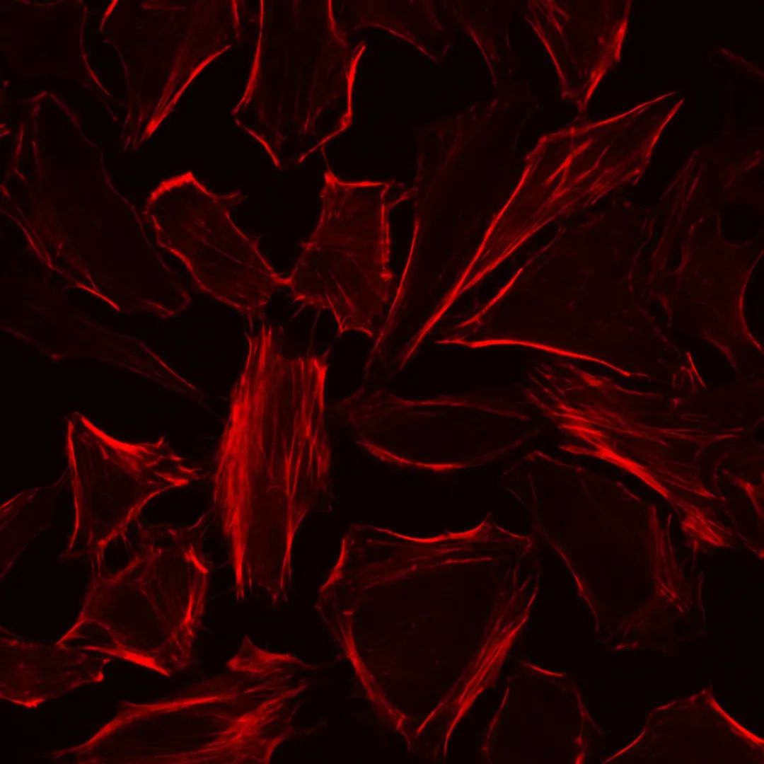
PI, or propidium iodide, is an analog of ethidium bromide that binds to DNA strongly. When it is embedded in double-stranded DNA, it releases red fluorescence to stain DNA or the nucleus. PI cannot penetrate the intact cell membrane, but it can penetrate the damaged cell membrane of late apoptotic cells and dead cells. To take account of this feature, PI is usually used in combination with fluorescent probes such as Calcein-AM or FDA to stain and observe dead cells, or flow cytometry is used for relative quantitative detection of apoptosis and cell cycle. The maximum excitation wavelength of PI-double-stranded DNA complex was 535nm, and the maximum emission wavelength was 615nm.
The PI staining solution of this product is ready-to-use with a concentration of 100 μg/mL, it can be directly used to stain the nuclei of necrotic cells or tissues, and the cell suspension can be used to detect cell cycle by flow cytometry after staining with this staining solution.
Storage and Handling Conditions
Wet ice transportation; Store at 2-8℃ away from light, valid for 6 months.
Component
| Component | G1021-10ML |
| PI Stain | 10 mL |
| Product Manual | |
Assay Protocol
l Flow Cytometry Assays For Cell Cycle Procedures:
1. The cultured viable cells were digested, washed with PBS, and collected by low-speed centrifugation to obtain cell precipitation.
2. Slowly add 1-3 mL of 90% ethanol precooled at 20 ° C to the cell precipitation, resuspended the cells, and ice bath overnight.
3. Cells were collected by centrifugation at 1500 for 5min, resuspended in PBS, and centrifuged again to remove the supernatant.
4. Cells were resuspended with 250 μL PBS.
5. Add 2 μL of RNase A (G3405 and G3413 are recommended) at a concentration of 1 mg/mL, and then water bath at 37℃ for 40 min.
6. Add 50 µL PI dye solution and incubate in the dark for 20 min (the time can be adjusted according to the staining results of experimental materials).
7. Flow cytometry detection.
ll Fluorescence Microscopy to Observe The Procedure of Living Dead Cells:
1. After cultured adherent cells, the culture medium was removed and washed twice with PBS.
2. The PI dye solution was diluted 10-20 times with PBS to prepare a final concentration of 5-10 μg/mL PI staining working solution. An appropriate amount of PI staining working solution was added to the well plate to cover the cells, and the cells were incubated for 5-10 min at room temperature in the dark.
3. The staining work was abandoned, and the cells were covered with PBS and observed by fluorescence microscope. Nuclei of dead or late apoptotic cells show red fluorescence.
Note
1. All fluorescent dyes are quenched, so it is recommended to complete the detection on the same day as possible after dyeing.
2. Wear a lab coat and disposable gloves during operation.
For Research Use Only!
Product recommendation
|
Cat.No.
|
Product Name
|
Spec.
|
Operation
|
|---|
|
G0002-2L
|
Phosphate Buffered Saline (PBS, Powder)
|
2 L
|
|
|
G1011-10ML
|
Hoechst 33258 Staining Solution (Ready-To-Use)
|
10 mL
|
|
|
G1012-10ML
|
DAPI Staining Reagent (Ready to Use)
|
10 mL
|
|
|
G1019-10ML
|
Trypan Blue Stain
|
10 mL
|
|
|
G1401-5ML
|
Anti-fade Mounting Medium
|
5 mL
|
|
|
G4101-200T
|
MTT Cell Viability Assay Kit
|
200 T
|
|
|
G4102-100T
|
Brdu Detection Kit
|
100 μL+30 mL×4
|
|
|
G4102-50T
|
Brdu Detection Kit
|
50 μL+30 mL×4
|