Description
Product Information
| Product Name | Cat.No | Spec. |
| Reactive Oxygen Species (ROS) Assay Kit | G1706-100T | 100T |
Introduction
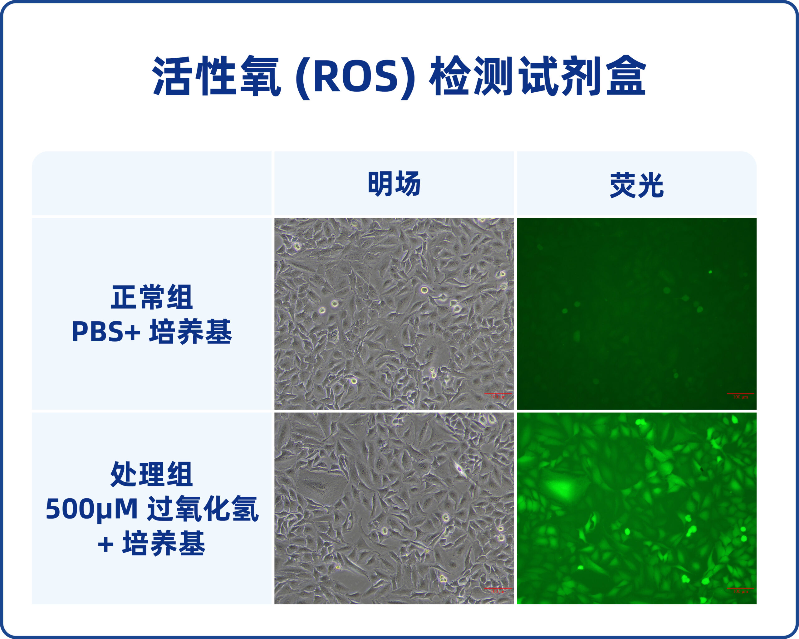
Reactive oxygen species (ROS) Assay Kit, also known as ROS Assay Kit, is a Kit for ROS detection based on the fluorescent probe DCFH-DA. DCFH-DA, also known as 2′,7′ -dichlorofluorescein diacetate, molecular weight 487.29, does not have fluorescence itself, can freely cross the cell membrane. After entering cells, DCFH can be hydrolyzed by esterase, which is widely existed in cells. DCFH is not permeable to cell membrane and thus is trapped inside the cell. Non-fluorescent DCFH can be oxidized by intracellular reactive oxygen species (ROS) to generate fluorescent DCF, and the intensity of fluorescence signal generated is proportional to the level of intracellular ROS. The level of intracellular reactive oxygen species can be indirectly evaluated by fluorescence microscopy, flow cytometry or laser confocal microscopy.
Reactive oxygen species (ROS) Assay kit can quickly and effectively detect intracellular ROS with high sensitivity and easy to use. At the same time, ROS positive induction reagent is provided, which can induce ROS levels in multiple types of cells in a short time to provide positive samples. Taking the amount of sample added to each well of the 6-well plate as the standard calculation, this kit can be measured about 100 times.
Storage and Handling Conditions
Transport with wet ice; Store at -20℃ away from light, valid for 12 months.
Components
| Component Number | Component | G1706-100T |
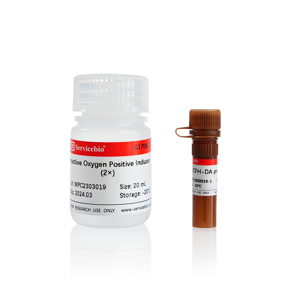
| G1706-1 | DCFH-DA probe | 100 μL |
| G1706-2 | Reactive Oxygen Positive Inducer (2×) | 20 mL |
| Instruction Manual | 1 pc | |
Usage
1. Preparation before experiment:
1.1. Preparation of DCFH-DA working solution: DCFH-DA working solution was prepared by diluting DCFH-DA probe with serum-free cell medium or Earle’s balanced salt solution (G4213) at a ratio of 1:1000.
1.2. (Optional) ROS positive inducer working solution preparation :1 × ROS positive inducer working solution was prepared by mixing and diluting the ROS positive inducer (2×) with serum-free cell medium or Earle’s balanced salt solution (G4213) in a 1:1 volume.
2. (Optional) Cell preparation for positive control group:
2.1. The original medium was removed by adherent cells in good condition, and the medium was removed by centrifugation;
2.2. Adherent cells or resuspended cells were covered with 1× reactive oxygen species positive inducer working solution and incubated in a CO2 incubator at 37℃ for 45 min under light(The sensitivity of different cells is different. If the increase of reactive oxygen species is not observed after 45 minutes of stimulation, the induction time can be appropriately prolonged or the concentration of inducer can be increased; if the increase of reactive oxygen species is too fast, the induction time or the concentration of inducer can be appropriately decreased).
3. Reactive oxygen species detection of adherent cells:
3.1. Cell seed plate: at least 1 day in advance, the cells in good condition are evenly seeded into the well plate according to a certain density, and drugs or other pretreatment is carried out according to the experimental needs (the inoculation density is determined by the cell size, growth rate and other factors, so as to ensure that the cell confluence is 50-70% during the test);
3.2. Cell washing: Remove the cell culture medium and wash it with PBS buffer (G4202) 1-2 times to reduce the interference of serum, drugs and other substances on the experimental results:
3.3. Loading probe: Remove the washing buffer by suction, add the corresponding volume of DCFH-DA working solution according to the table below, and incubate in a CO2 incubator at 37℃ for 30 min under light. Then the DCFH-DA working solution was aspirated and washed with PBS buffer 2-3 times to fully remove the excess probe; Finally, the cells were covered with PBS.
| Cell Culture Plate | Reactive Oxygen Species Detection Working Solution/Well |
| 6-well | 1000 μL |
| 12-well | 500 μL |
| 24-well | 250 μL |
| 48-well | 200 μL |
| 96-well | 100 μL |
3.4. ROS detection: The labeled cells in the well plate were directly placed into the fluorescence microscope or confocal microscope for observation. Or after digestion, the cells were collected and detected by fluorescence spectrophotometer, microplate reader, flow cytometer and other instruments. With the excitation wavelength of 488nm and emission wavelength of 525nm, the fluorescence spectrum of DCF is very similar to that of FITC, and the parameters of FITC can be set to detect DCF.
2. Detection of reactive oxygen species in suspension cells (also applicable to adherent cells resuspended after digestion and pretreatment):
2.1. Cell pretreatment: Select cells in good condition according to the experimental requirements, and centrifuge them at 1000 RPM for 3-5 min after drug or other pretreatment according to the experimental requirements to collect cells (cell density shall be determined according to the subsequent detection system, method and total amount, for example, the number of cells in a single sample by flow detection shall be no less than 104);
2.2. Cell washing: Wash 1-2 times with PBS buffer, centrifuge at 1000 RPM for 3-5 min to remove supernatant and collect cells;
2.3. Probe loading: Add a certain volume of DCFH-DA working solution to make the cell density of 1×10⁵-5×10⁵ cells /mL, and incubate in a CO2 incubator at 37℃ for 30 min in the dark. During the period, gently shake the mixture every 5 min to ensure full contact between the probe and the cell. At the end of the incubation, the DCFH-Da working solution was removed by centrifugation at 1000 RPM for 3-5 min and washed 2-3 times with PBS buffer to adequately remove excess probes. Finally, the cells were resuspended in PBS.
2.4. ROS detection: Cells labeled with probe can be directly detected by fluorescence spectrophotometer, microplate reader, flow cytometer, etc. It can also be dropped onto a slide and observed with instruments such as fluorescence or confocal microscopy. With the excitation wavelength of 488nm and emission wavelength of 525nm, the fluorescence spectrum of DCF is very similar to that of FITC, and the parameters of FITC can be set to detect DCF.
Note
1. If the overall fluorescence intensity after probe labeling is too high or too low during the experiment, the dilution ratio and incubation time of DCFH-DA probe can be adjusted appropriately.
2. If the probe does not enter the cell, it will cause a high background. Please clean it as far as possible.
3. The positive inducer of reactive oxygen species is mainly used for the induction of positive control samples. According to the experimental needs, whether to be a positive control or not.
4. Each centrifugation of cells will cause a certain loss to the total cell density. Please pay attention to control the initial cell amount to prevent the final cell amount from being insufficient for detection.
5. Due to quenching of fluorescent probe, please try to shorten the time from probe incubation to detection (except stimulation time).
6. For your health and safety, please wear laboratory coat and gloves when operating.
For Research Use Only!

