No products in the cart.


40. Booking other animal brain Microtome Blade, price can be discussed.
$1.00
In Stock
Description
Product Description:
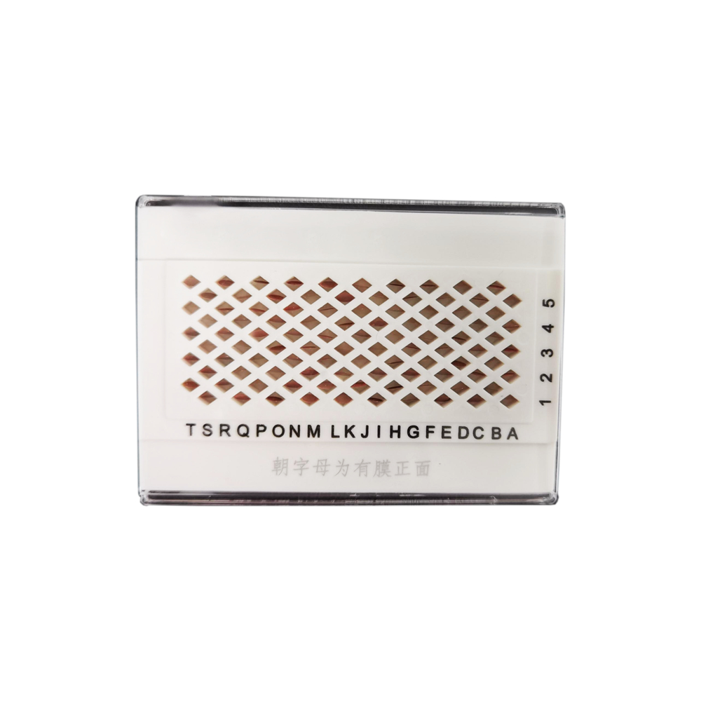
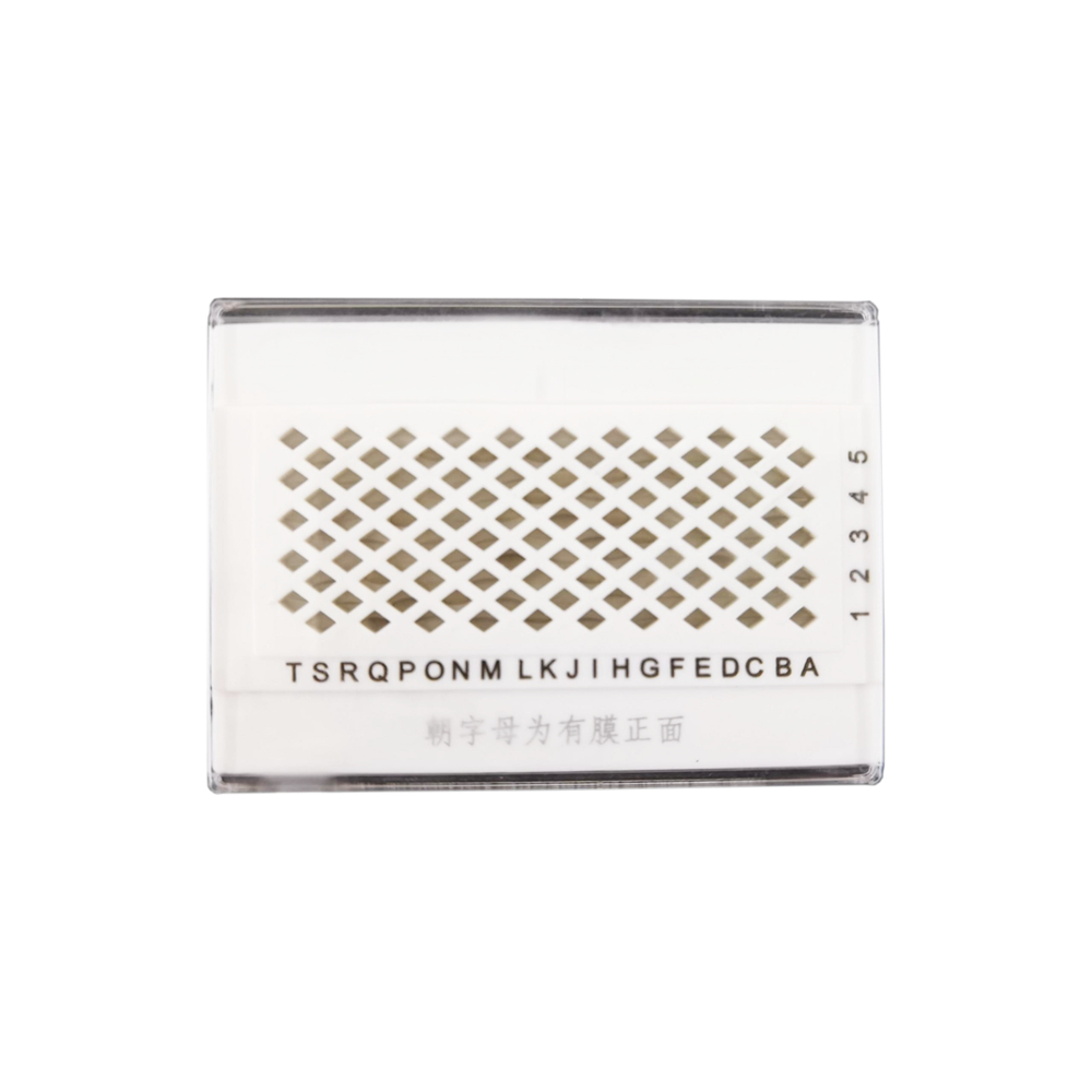
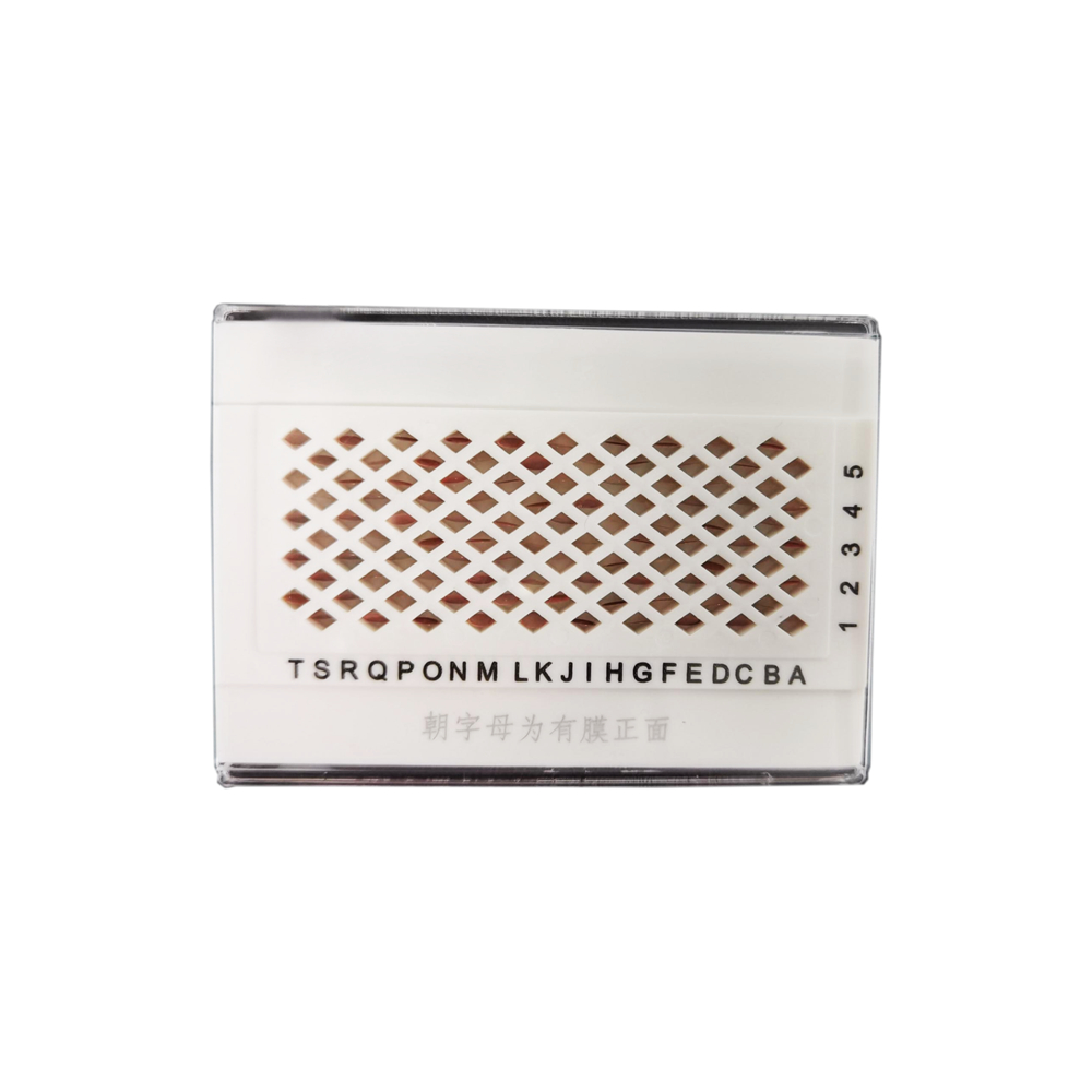
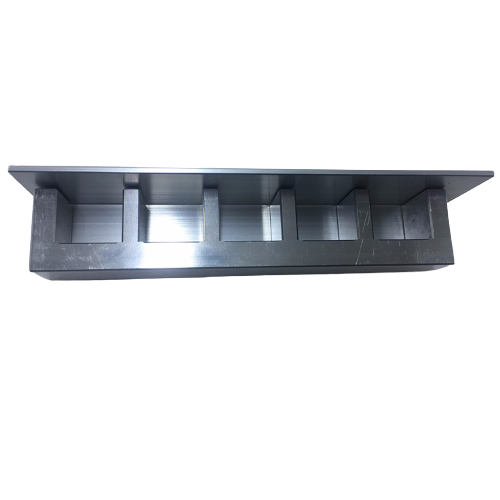
The animal brain slicing mold is primarily used as a tool for cutting and preparing specific brain sections from animal brain tissues. Our company provides two forms of brain slicing molds for rat and mouse brains: coronal sections, which are perpendicular to the brain midline, and sagittal sections, which are parallel to the brain midline. These molds are made of high-quality aluminum alloy material, ensuring precise dimensions, smooth cutting edges, and consistent thickness, guaranteeing reproducibility during the slicing process. We also accept customized orders for brain slicing molds for other animal species such as monkeys, pigs, chickens, ducks, rabbits, and guinea pigs.
| Name | Catalog Number | Description | Animal | Sectioning Direction | Slice Thickness (mm) |
|---|---|---|---|---|---|
| Slice Mold | SQP-H12 | Mouse Brain, Coronal | Mouse | Coronal | 1mm |
| Slice Mold | SQP-H23 | Rat Brain, Coronal | Rat | Coronal | 1mm |
Product Features:
- Made of high-quality aluminum alloy material, resistant to high temperatures, easy to clean, and durable.
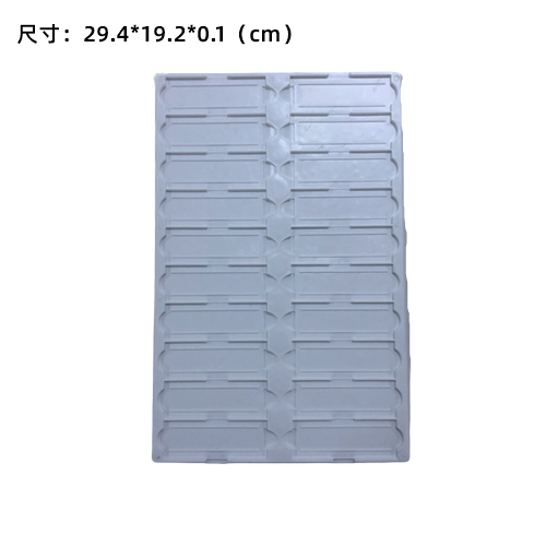
- Digitally engraved markings for easy positioning, with 0.35mm wide grooves and 1mm spacing.
- Provides two specifications: coronal slice mold (perpendicular to the brain midline) and sagittal slice mold (parallel to the brain midline).
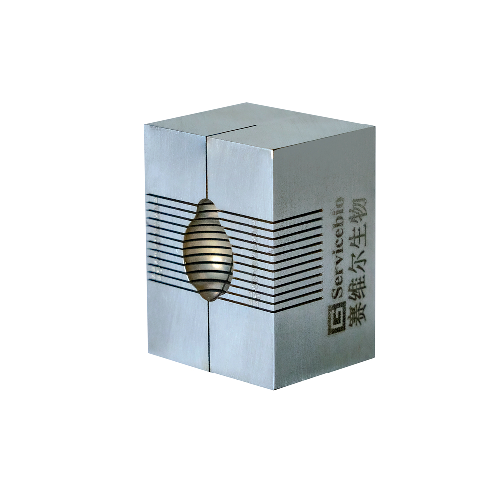
- The coronal brain mold has a sagittal midline for easy separation of the left and right brain hemispheres.
Applicable Fields:
- Used for preliminary positioning of fixed brain tissue, facilitating fine slicing for paraffin embedding, frozen sections, vibratome sections, and ultra-thin sections.
- Used for the precise positioning of fresh brain tissue, combining with brain atlases for preliminary localization of neural nuclei and specific brain regions. It allows the extraction of targeted tissue samples for protein, molecular, biochemical, and other analytical studies, such as the analysis of neurotransmitter and metabolite concentrations.
Instructions for Use:
- Place the dissected or fixed animal brain tissue into the corresponding mold groove based on the required sectioning direction and tissue shape. Ensure that the base of the olfactory bulb of the tissue is aligned with the base of the olfactory bulb on the mold for better positioning.
- Determine the desired sectioning location and insert two paraffin blades (recommended using Saville BioTech WDP819 Leica blades or WBL04-LR35 ERMA paraffin blades) into the slicing grooves on both sides of the target tissue. Press both ends of the blades simultaneously to cut the tissue. Extract the cut brain section gently with forceps for further experiments.
- Remove the remaining tissue from the groove, wash the mold with water, and let it air dry for future use.
Suggested Brain Regions for Common Slice Positions:
For mice weighing 20-30g, coronal brain sections (aligning the base of the olfactory bulb of the tissue with the base of the olfactory bulb on the mold) are recommended.
| Brain Region | Corresponding Range on Mold | Slice Position on Mold | Fine Slicing for Paraffin/Frozen/Vibratome |
|---|---|---|---|
| Olfactory bulb | 1-2 | Start from line 2 | From the top of line 1 |
| Frontal cortex | 3-4 | Cut lines 3 and 4 | Start from line 3 |
| Striatum | 4-5 | Cut lines 4 and 5 | Start from line 4 |
| Amygdala | 5-6 | Cut lines 5 and 6 | Start from line 5 |
| Dorsal Hippocampus, Thalamus, Hypothalamus | 6-7 | Cut lines 6 and 7 | Start from line 6 |
| Ventral Hippocampus, Substantia Nigra | 6-7 | Cut lines 6 and 7 | Start from line 7 |
| Cerebellum | 9-11 | Cut lines 9 and 11 | Start from line 9 |
Note: The intersection of line 4 with the sagittal midline represents the approximate position of the anterior commissure. For other specific brain regions, refer to the mouse brain atlas for preliminary localization.
.