No products in the cart.


4. Protein Free Rapid Blocking Buffer (+Western Blot Common Issues Reference Table), 500 ml $149
$149.00
In Stock
Description
Product Introduction Related Products
Product Information
| Product Name | Cat.No. | Spec. |
| Protein Free Rapid Blocking Buffer | G2052-500ML | 500 mL |
Description/Introduction
This product is a new generation of protein-free fast sealing liquid, which is a mixed liquid of various polymers. It can quickly fill the voids on the surface of solid support and reduce the interference of non-specific signals; The overall sealing effect is obviously better than the traditional sealing solution based on BSA (bovine serum albumin), skimmed milk powder, casein, etc; It can be used for blocking and primary antibody dilution in Western blot (WB), ELISA, IHC, if and other experiments.
Fast and efficient: the blocking time is only 5 minutes, and the signal-to-noise ratio is stronger than that of BSA, skim milk powder, casein and other traditional blocking agents.
Low background: this blocking solution does not contain serum, albumin and other proteins, ensuring a high signal-to-noise ratio.
Good compatibility: This product is compatible with horseradish peroxidase (HRP), alkaline phosphatase (AP) and biotin labeled secondary antibodies; No preservatives affecting the activity of HRP and AP; Since it does not contain biotin, it will not interfere with the detection based on biotin.
Easy to use: This product contains tbst and can be directly used for dilution of primary antibody.
Storage and Handling Conditions
Wet ice bag transportation; Stored at 4℃;valid for 12 months;It can be stored at – 20 ℃ if not used for a long time.
Component
| Component | G2052–500ML |
| Protein Free Rapid Blocking Buffer | 500 mL |
| Manuals | One copy |
Assay Protocol / Procedures
1. Membrane blocking in Western blot
a) Wash with tbst for 1-2 minutes after the completion of film transfer;
b) According to the size of the membrane, select a suitable container, add a certain volume of protein free fast sealing liquid to ensure that the sealing liquid can completely cover the membrane, and place it on the pendulum or horizontal shaking table for 5 minutes;
c) The primary antibody was diluted with protein-free rapid blocking solution, and subsequent incubation could be carried out after blocking.
2. Blocking of substrate in ELISA experiment
a) ELISA plates were coated with antigen or antibody;
b) Add 300 per hole μ L protein-free fast blocking solution and incubated at 37 ℃ for 5 minutes;
c) According to the requirements of ELISA experiment, continue the following steps such as washing or plate throwing.
3. blocking in IHC and if experiments
a) In IHC and if experiments, this reagent is used as a solvent to Dissolve 3% BSA. Compared with BSA dissolved in PBS, it has stable sealing performance and excellent sealing effect in various tissues; For antibodies with good specificity, this reagent can be directly used as a blocking agent
b) After the tissue sections are prepared, add protein-free blocking agent or BSA dissolved protein-free blocking agent, and stand at room temperature for 30 minutes (with tissue difference); The sample must be completely covered to avoid high background caused by sample drying
Note:
1. No one blocking agent can be applied to all antigen and antibody detection systems. In case of antibodies unsuitable for this reagent, please replace other types of blocking agents such as BSA or skim milk powder.
2. After use, store the remaining solution at 2-8 ℃ to avoid pollution.
3. The reagent has a certain viscosity, and the pipette needs to be slowly sucked and slowly released to ensure the accuracy of the volume.
4. This product is recommended to be used only once, and repeated use may lead to the decrease of sealing effect; For antibodies with good specificity and high signal-to-noise ratio, the blocking solution can be reused; Do not mix the recovered sealing liquid with the unused sealing liquid.
5. For your safety and health, please wear experimental clothes and disposable gloves.
For Research Use Only!
Ver. No.: V1.0-202208
Product recommendation
|
Cat.No.
|
Product Name
|
Spec.
|
Operation
|
|---|
|
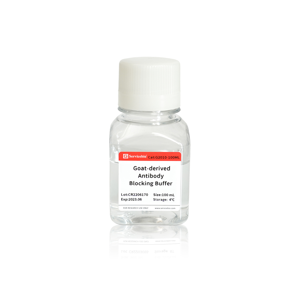
G2010-100ML
|
Blocking Buffer (Special Use for Goat-Derived Antibody)
|
100 mL
|
|
|
G2052-500ML
|
Protein Free Rapid Blocking Buffer
|
500 mL
|
Western Blot Common Issues Reference Table:
Problem
Possible Causes
Solution
- No signal
- Insufficient active HRP on PVDF or NC membrane
- Mismatched species source between primary and secondary antibodies
- Poor recognition or low potency of the primary antibody
- Unsuccessful transfer of proteins
- Perform ECL secondary incubation or extend the incubation time between ECL and the membrane
- Switch to a more sensitive ECL detection system
- Increase the sample amount
- Increase the concentration of the primary antibody or switch to a higher potency antibody. Establish a positive control to ensure the antibody recognizes the antigen.
- Ensure the secondary antibody matches the species source of the primary antibody
- Suggest running a protein marker during electrophoresis and confirm successful protein transfer
- Rapid signal decay
- Deteriorated ECL reagent
- Excessive active HRP on PVDF or NC membrane
- Switch to a fresh batch of ECL reagent
- Decrease the sample amount
- Decrease the concentration of the primary antibody
- Decrease the concentration of the secondary antibody
- High background
- Cross-reactivity between blocking agent and related reagents
- Inadequate washing steps
- Prolonged exposure time
- Insufficient blocking
- Switch to a protein-free rapid blocking solution
- Extend the washing time, especially for 0.22 µm PVDF membranes
- Shorten the exposure time or switch to a lower sensitivity ECL detection system
- Extend the blocking time
- Brown/yellow bands or ghost bands
- Excessive active HRP on PVDF or NC membrane, resulting in accumulation of oxidized and inactivated HRP that appears as visible brown/yellow bands
- Decrease the sample amount
- Decrease the concentration of the primary antibody
- Decrease the concentration of the secondary antibody
- Non-specific bands
- Related to the antibody, generally fewer non-specific bands with monoclonal antibodies compared to polyclonal antibodies
- Sample degradation
- Choose antibodies with high specificity and potency
- Re-extract fresh samples
- Unstained white spots within bands
- Bubbles between filter paper and gel, gel and membrane, or membrane and filter paper during transfer
- Ensure that bubbles are completely removed during the transfer process
- Deformed bands
- Inhomogeneous gel formation during gel casting
- High temperature during electrophoresis causing gel deformation
- High temperature during transfer causing gel deformation
- Excessive tightness of the transfer clamp causing gel squeezing and deformation
- Ensure homogeneous gel formation, especially at the interface between upper and lower gel layers
- Lower the voltage during electrophoresis to reduce heat generation or perform electrophoresis in a cold bath
- Perform the transfer process in a fully chilled condition
- Adjust the tightness of the transfer clamp to provide moderate pressure
- Blocking of the substrate in ELISA
a) Coat the ELISA plate with antigen or antibody;
b) Add 300 μL of protein-free rapid blocking solution per well and incubate at 37°C for 5 minutes;
c) Proceed with subsequent steps such as washing or adding the subsequent reagents according to the requirements of the ELISA assay.
- Blocking in IHC and IF experiments
a) Use this reagent to dissolve BSA at 3% (mass/volume ratio) for IHC and IF experiments. Compared to BSA dissolved in PBS, this reagent provides stable blocking performance and excellent blocking effect in various tissues. For highly specific antibodies, this reagent can be directly used as the blocking agent.
b) After preparing tissue sections, add protein-free rapid blocking solution or BSA-containing protein-free rapid blocking solution and incubate at room temperature for 30 minutes (depending on tissue variations). Ensure complete coverage of the sample to prevent sample drying and high background.
Precautions:
- No single blocking agent is suitable for all antigen and antibody detection systems. If this reagent is not suitable for a specific antibody, consider using other types of blocking agents such as BSA or skim milk.
- After use, store the remaining solution at 2-8°C to avoid contamination.
- This reagent has certain viscosity. When using a pipette, aspirate and dispense slowly to ensure accurate volume.
- This product is recommended for single use only. Repeated use may result in reduced blocking efficiency. For antibodies with high specificity and signal-to-noise ratios, the blocking solution can be reused. Do not mix recycled blocking solution with unused solution.
- For your safety and health, please wear laboratory attire and disposable gloves during operation.
This product is for research purposes only and is not intended for clinical diagnosis.
Version: V1.0-202205
(Product packaging is being upgraded, refer to the actual product.)