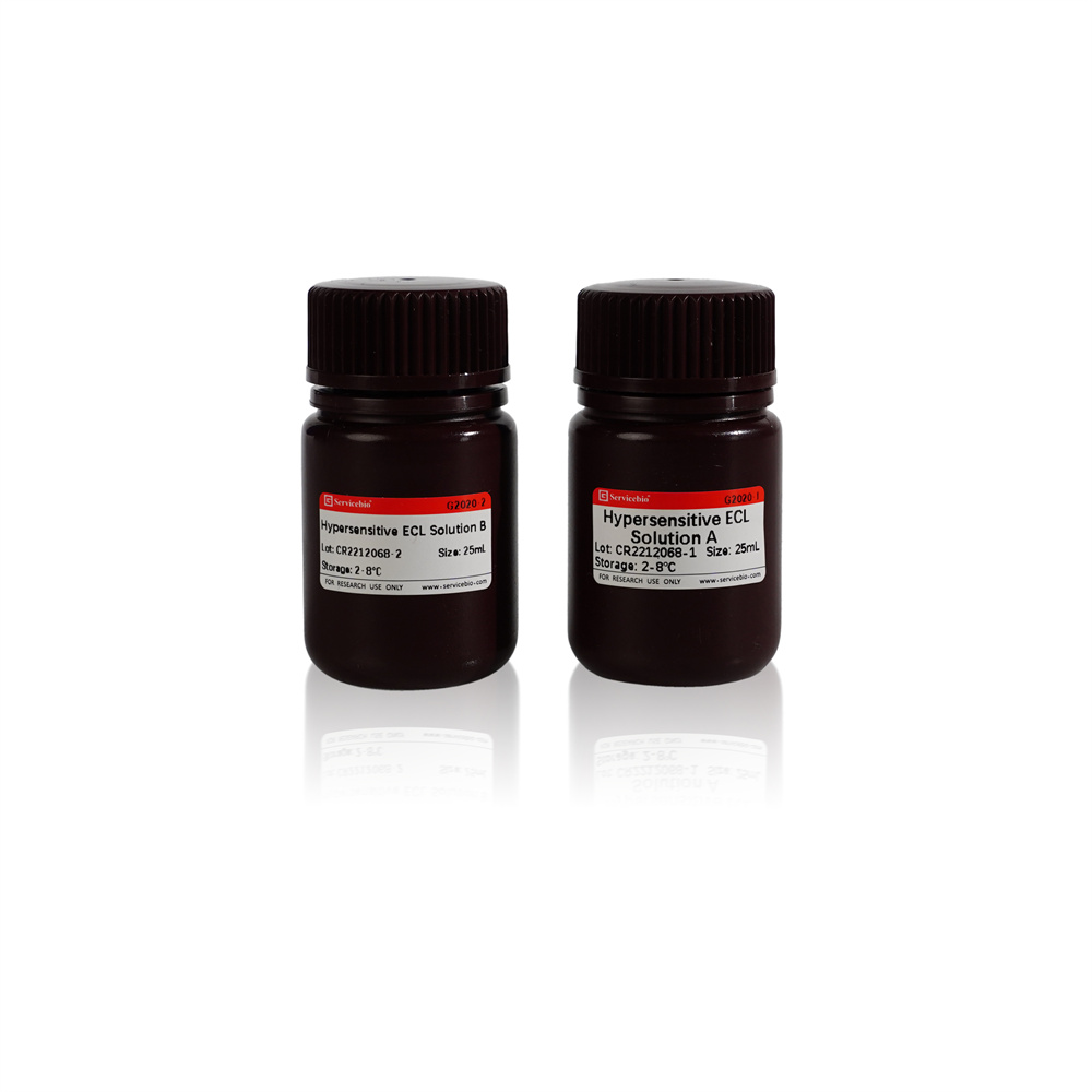
Description
Please see the videos for the relative products
Product Information
| Product Name | Cat. No. | Spec. |
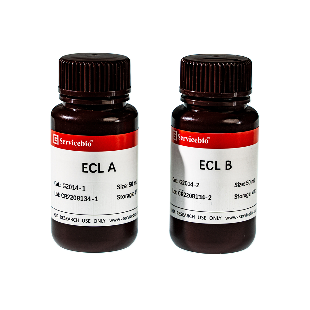
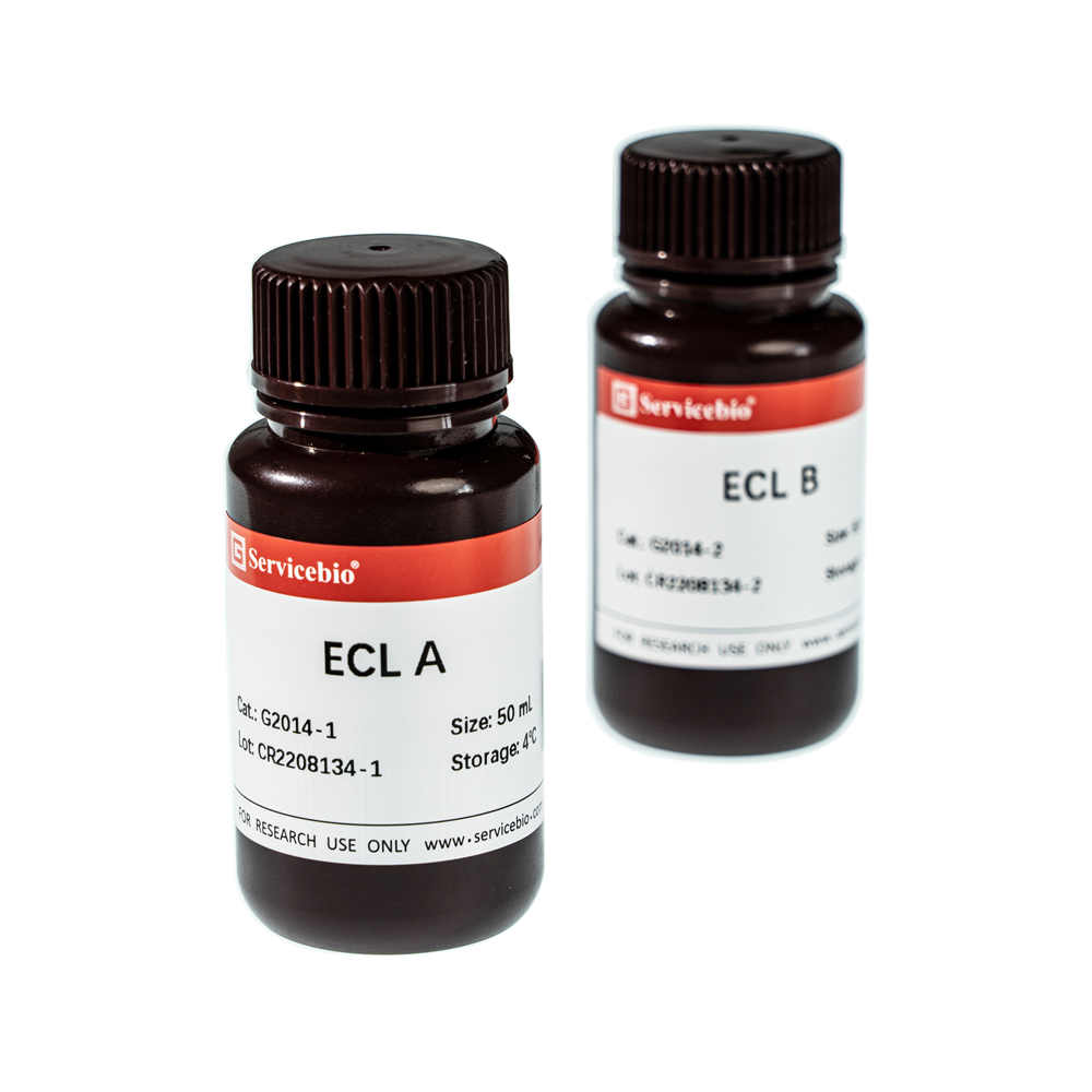
| ECL Chemiluminescence Kit | G2014-100ML | 2×50 mL |
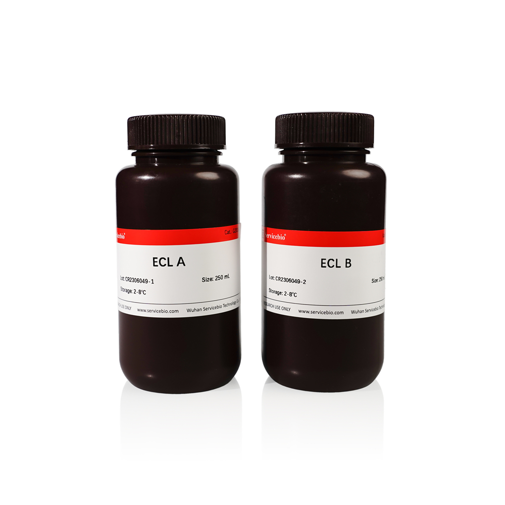
| G2014-500ML | 2×250 mL |
Product Description/Introduction
The ECL chemiluminescence kit is a highly sensitive chemiluminescence kit based on the principle of Luminol ECL chemiluminescence. This product can chemically react with horseradish peroxidase (HRP) coupled with secondary antibodies to produce fluorescence, which can be detected by X-ray tablet pressing or other appropriate fluorescence imaging equipment (CCD camera, etc.).
Storage and Shipping Conditions
Ship with wet ice; Store in the dark at 2-8℃; valid for 12 months.
Assay Protocol/Procedures
1. Preparation of ECL working solution: Mix ECL solution A and ECL solution B in equal volume, and store in the dark at 4℃. Use it within 2 days.
2. In the Western blot experiment, PVDF membrane is incubated by the secondary antibody, washed for several times, and the excess liquid is absorbed by the filter paper. Two layers of PE gloves or other transparent films are affixed to the exposure box. The PVDF membrane protein is placed face up between the two layers of the exposure box. The mixed ECL working solution is added to cover the film and placed on the film for 1-2 min.
3. Absorb the ECL working fluid with filter paper or blotting paper, cover the upper film and start pressing film.
4. The pressed film is developed and fixed with developing and fixing reagents (G2019, G2023 and G2024 are recommended). Adjust the exposure conditions according to the luminous intensity.
Note
1. The pipette tips must be replaced during the liquid transferring process of ECL liquid A and liquid B. Cross contamination of liquid A and liquid B will lead to the gradual failure of liquid A or B. In addition, the contamination of metal ions will reduce the sensitivity of this reagent. Please pay attention to use clean pipette tips, Seal well after use.
2. If the background after exposure is very deep, the reason may be that the concentration of the secondary antibody or the primary antibody is too high, or the sealing solution is not suitable, and other sealing solution should be used.
3. If the fluorescence quenches rapidly, it may be due to the over-strong fluorescence of the target band, resulting in the rapid consumption of ECL by HRP.
4. If there is no luminous signal, the target protein may be weakly expressed and the compression time may be extended.
5. For your safety and health, please wear safety glasses, gloves, or protective clothing.
For Research Use Only!
|
Cat.No.
|
Product Name
|
Spec.
|
Operation
|
|---|
|
G0001-2L
|
Tris Buffered Saline (TBS,Powder)
|
2 L
|
|
|
G2001-250ML
|
Coomassie (Bradford) Protein Quantitative Assay Kit
|
250 mL
|
|
|
G2003-50T
|
SDS-PAGE Gel Preparation Kit
|
50 T
|
|
|
G2004-100ML
|
30% Acrylamide-Bisacrylamide (29:1)
|
100 mL
|
|
|
G2005-100ML
|
40% Acrylamide-Bisacrylamide (37.5:1)
|
100 mL
|
|
|
G2007-1ML
|
Phosphoprotease Inhibitor
|
1 mL×2
|
|
|
G2009-100ML
|
Western Secondary Antibody Dilution Buffer
|
100 mL
|
|
|
G2011-100ML
|
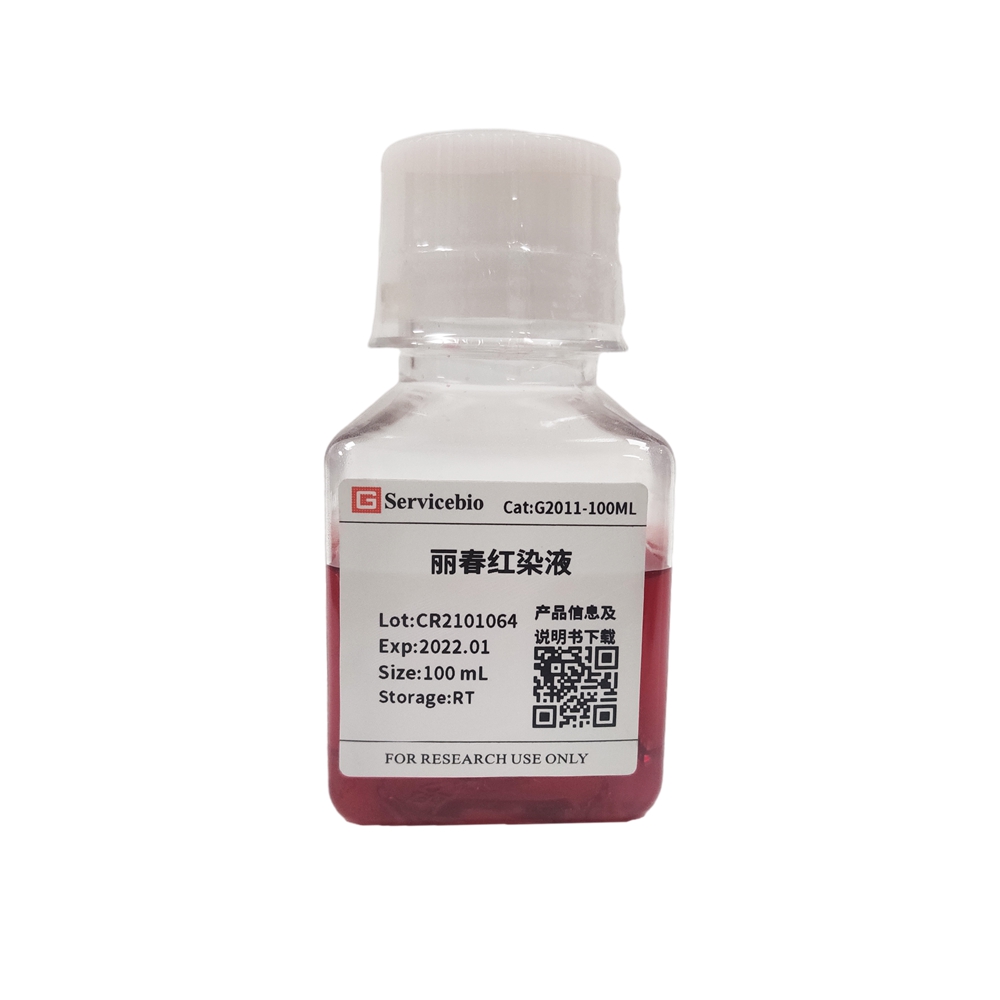
Ponceau S Staining
|
100 mL
|
|
|
G2012-250ML
|
Coomassie Bright Blue Dye Kit
|
250mL+500mL
|
|
|
G2017-1L
|
Tris-Glycine Transfer Buffer (Powder)
|
1 L
|
|
|
G2018-1L
|
Tris-Glycine SDS-PAGE Running Buffer (Powder)
|
1 L
|
|
|
G2020-50ML
|
Hypersensitive ECL Chemiluminescence Kit(FG)
|
25 mL×2
|
|
|
G2025-100ML
|
Primary Antibody Diluent
|
100 mL
|
|
|
G2026-200T
|
BCA Protein Quantitative Detection Kit
|
200 T
|
|
|
G26616-250UL
|
Protein Marker 10-180 KDa
|
250 μL
|
|
|
G26619-250UL
|
Protein Marker 10-250 kDa
|
250 μL
|
|
|
G6002
|
Transfer Sponge
|
2 pcs/box
|
|
|
G6007-50
|
Transfer Membrane Filter Paper (thin)
|
50 pcs/bag
|
|
|
G6015-0.45
|
PVDF Membrane (0.45 μm)
|
0.45 μm 13.25*15cm
|
|
|
G6015-1-0.22
|
PVDF Membrane (0.2μm)
|
0.2 μm 13.25*15cm
|
|
|
G6051-1.0T
|
Glass Plate Set, 1.0 mm (for Liuyi,with reference line)
|
1.0 mm
|
|
|
WGA0018
|
Cassette
|
10.4*13 Inch
|

