Description
Product Information
| Product Name | Cat.No. | Spec. |
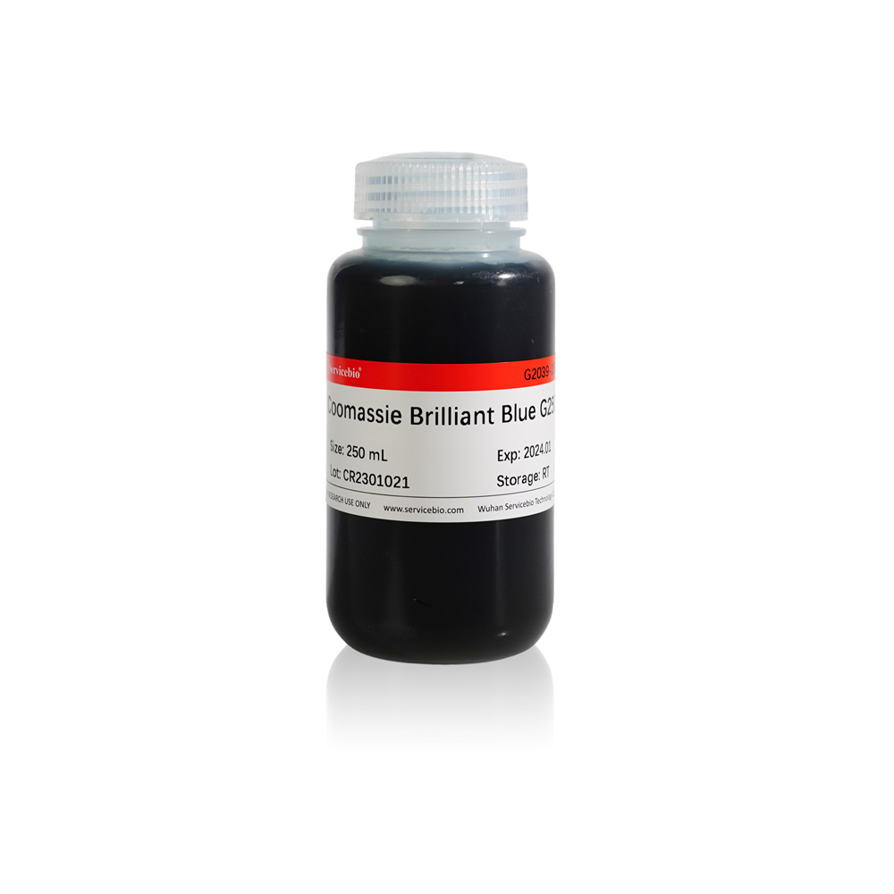
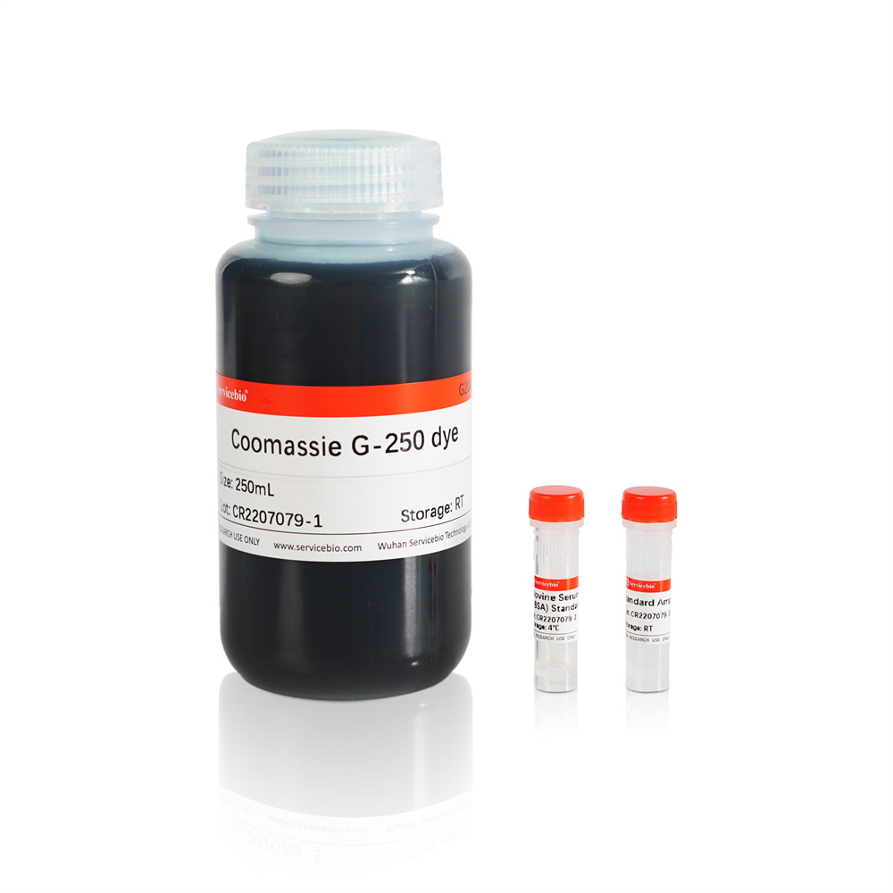
| Coomassie Brilliant Blue G250 | G2039-250ML | 250 mL |
Description
Coomassie Brilliant Blue G250 binds proteins rapidly. Coomassie brilliant blue G250 is red in the free state, and its maximum optical absorption is at 488 nm, and it has a maximum absorption value at 595 nm after binding to protein, and the optical absorption value is proportional to the protein content, so it can be used for the quantitative detection of protein. This product can also be used as a supplement to the G2001 Bradford Assay protein quantitative detection kit.
Storage and Handling Conditions
Store and transport at room temperature, valid for 12 months
Assay Protocol
1. (Optional) Drawing standard curve (microplate reader method) : Dissolve BSA in water to prepare 0.5 mg/mL protein standard working solution. The protein standard working solution was added to the 96-well plate at levels of 0,1,2,4,8,12,16,20 μL, and then the gradient working solution was supplemented to 20 μL with PBS or saline. The gradient curves of protein concentration were 0, 25, 50, 100, 200, 300, 400 and 500 μg/mL. If only relative quantitative comparisons are made, no standard curves are required.
2. Prepare the sample to be tested: The protein sample to be tested is diluted appropriately (the protein concentration of the sample can be detected by pre-experiment to be within the range of the standard curve, to ensure the reliability of the test result), and 20 μL of each sample is added to the 96-well plate. The sample to be tested should be diluted in the same solution as the protein standard.
3. Detection: 200 μL Coomassie brilliant blue G250 solution was added to each well and thoroughly mixed (the 96-well plate could be placed on the oscillator for 30 s). After 3-5 min at room temperature, the standard curve No. 0 was used as a reference, and the colorimetric measurement was performed at 595 nm wavelength, and the absorbance value of each well was recorded.
4. Calculation: The gradient protein content (μg/mL) in the standard curve was taken as the abscissa, and the light absorption value was taken as the ordinate to draw the standard curve. According to the absorbance value of the sample, the protein concentration of the sample to be measured in the corresponding well can be found on the standard curve (μg/mL), and then multiplied by the dilution of the sample, the actual protein concentration of the sample to be measured.
In addition: if the spectrophotometer is used to determine, the glass test tube or glass colorimetric tube is used as the reaction vessel to make standard curves. After proper dilution of the protein sample to be tested, add it to a new glass test tube or colorimetric tube with a sample size of 1 mL. Add 3 mL of Coomassie brilliant blue G250 solution to the standard curve gradient tube and sample tube, mix thoroughly, stand at room temperature for 3-5 min, then use spectrophotometer for colorimetric detection.Detection: The wavelength of the spectrophotometer was set to 595 nm, and the standard curve and the sample to be tested were zeroed with the standard curve No. 0 tube as the reference. Standard curves were drawn as described in Step 4 and protein concentrations in the samples to be tested were calculated.
Note:
1. The product should be restored to room temperature before use, and mixed upside down, so as not to affect the sensitivity of detection.
2. For your safety and health, please wear a lab coat and disposable gloves when operating.
For Research Use Only!
|
Cat.No.
|
Product Name
|
Spec.
|
Operation
|
|---|
|
G2002-100ML
|
RIPA Lysis Buffer (Strong)
|
100 mL
|
|
|
G2002-30ML
|
RIPA Lysis Buffer (Strong)
|
30 mL
|
|
|
G2003-50T
|
SDS-PAGE Gel Preparation Kit
|
50 T
|
|
|
G2004-100ML
|
30% Acrylamide-Bisacrylamide (29:1)
|
100 mL
|
|
|
G2006-250UL
|
50×Cocktail Protease Inhibitor
|
250 μL
|
|
|
G2007-1ML
|
Phosphoprotease Inhibitor
|
1 mL×2
|
|
|
G2008-1ML
|
PMSF (100mM)
|
1 mL
|
|
|
G2018-1L
|
Tris-Glycine SDS-PAGE Running Buffer (Powder)
|
1 L
|
|
|
G2033-100ML
|
RIPA Lysis Buffer (Weak)
|
100 mL
|
|
|
G2033-30ML
|
RIPA Lysis Buffer (Weak)
|
30 mL
|
|
|
G2053-100ML
|
1.5 M Tris-HCl (pH 8.8)
|
100 mL
|
|
|
G2054-100ML
|
1 M Tris-HCl (pH 6.8)
|
100 mL
|
|
|
G2055-5ML
|
10% SDS Solution
|
5 mL
|
|
|
G6019-9
|
Antibody Incubation Box (9 Grids)
|
9 grids
|
|
|
G6020-9
|
Antibody Incubation Box (9 Grids Light-Proof)
|
9 Grids Light-Proof
|
|
|
G6025-2
|
Antibody Incubation Box (2 Grids)
|
2 grids
|
|
|
G6026-4
|
Antibody Incubation Box (4 Grids)
|
4 grids
|


