No products in the cart.
")
3. Brdu Detection Kit. 100T $688 ( buy more get for big discount), 100 μL+30 mL×4 (BrdU)
$688.00
In Stock
Description
Product Information
| Product Name | Cat.No. | Spec. |
| Cell Counting Kit-8 (CCK-8 Kit) | G4102-50T | 50T |
| Cell Counting Kit-8 (CCK-8 Kit) | G4102-100T | 100T |
Description/Introduction
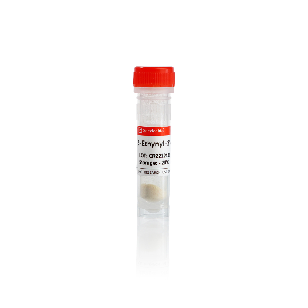
This product BrdU detection kit is used for cell proliferation detection of cells and tissue sections. BrdU (5-bromo-2-deoxyuridine), i.e. 5-bromodeoxyuridine, is a thymidine (T) analog. The principle of this kit is that BrdU can be incorporated into DNA synthesis phase (S phase) through competition instead of thymidine t. When the cells in vigorous division are mixed with BrdU, the cells containing BrdU can be detected by using anti BrdU antibody and antibody ligase or fluorescein as the indicator system. Combined with other cell markers for double labeling, the type and proliferation rate of proliferating cells can be determined, which is of great significance to the study of cell dynamics. BrdU enters tissue cells when injected in vivo or cultured in cells. The location of the incorporated DNA is accurate, which can effectively reflect the level of newly synthesized DNA in S-phase cells. Moreover, it is less affected by the internal and external environment of the cells, and the label will not be lost.
Storage and Handling Conditions
Wet ice transport; Membrane breaking solution, blocking solution (3% BSA) and 1 M HCl can be stored at 4 ℃, 4% paraformaldehyde can be stored at room temperature, and Anti-BrdU (mouse mAb) needs to be stored at – 20 ℃, with a validity period of 12 months.
Component
| Component Number | Component | G4102–50T | G4102–100T |
| G4102-1 | Membrane breaking solution | 30 mL | 30 mL |
| G4102-2 | blocking solution (3% BSA) | 30 mL | 30 mL |
| G4102-3 | 4% paraformaldehyde | 30 mL | 30 mL |
| G4102-4 | 1 M HCl | 30 mL | 30 mL |
| G4102-5 | Anti-BrdU (mouse mAb) | 50 uL | 50 uL |
| Protocol | 1 PICS | ||
Preparation
1. Prepare PBS buffer (recommended g4202), secondary antibody, gradient ethanol, sealing agent, etc;
2. Prepare the nucleus staining solution, and g1004 hematoxylin staining solution or g1012 DAPI staining solution (ready to use type) is recommended;
3. Preparation of anti BrdU primary antibody working solution: dilute anti BrdU (mouse mAb) 100 times with blocking solution (3% BSA) to prepare anti BrdU primary antibody working solution, which is ready for use and stored at 4 ℃;
4. Secondary antibody working solution: prepare the anti mouse secondary antibody labeled with HRP (followed by white light detection, matched with DAB color development kit, g1212 is recommended) or fluorescence (followed by fluorescence detection) according to the detection needs, and dilute it with PBS to prepare the secondary antibody working solution.
Assay Protocol / Procedures
Cell samples:
a. Cell preparation: prepare cell climbing pieces in advance to ensure normal cell state and good adhesion;
b. BrdU Incubation: BrdU containing cells are incubated in the incubator for a certain time in the absence of light. BrdU concentration and cell incubation time are different according to cell types. It is suggested to explore the best conditions in the pre experiment. The experimental conditions of the tested cells in the precautions are available for reference;
c. Cell fixation: the above cells incubated with BrdU were washed three times with PBS for 5 min each time. Add 1 ml of 4% paraformaldehyde to the well plate to cover the cells, and fix at room temperature for 20-30 min. Wash 3 times with PBS for 5 min each time;
d. Permeabilization: add membrane breaking solution to the well plate to cover the cells for 8-10 min, and wash with PBS for 3 times for 5 min each time;
e. Nuclear staining (optional):
In case of subsequent white light detection, the nuclei were stained with hematoxylin: add hematoxylin dye solution to the well plate for 5 min at room temperature, suck and discard hematoxylin dye solution, and wash with PBS for 3 times for 5 min each time;
In case of subsequent fluorescence detection, the nuclei were stained with DAPI: add DAPI dye solution to the well plate for 5 min at room temperature, suck and discard DAPI dye solution, and wash with PBS three times for 5 min each time;
f. Re fixation: add 4% paraformaldehyde dropwise to the well plate, incubate at room temperature for 10 min, and wash with PBS three times for 5 min each time;
g. Acidification: add 1 M HCl to the well plate to cover the cells, treat them at 37 ℃ for 10 min, and wash them with PBS three times for 5 min each time;
h. Post fixation: add 4% paraformaldehyde to the well plate again, incubate for 10 min at room temperature, and wash with PBS three times for 5 min each time;
i. Blocking: add blocking solution (3% BSA) to the well plate and incubate at room temperature for 30 min. Suck and discard the sealing liquid, and do not clean it;
j. Anti-BrdU antibody incubation: add anti BrdU primary antibody working solution to the well plate to cover the cells, and incubate overnight at 4 ℃. Suck and discard the anti BrdU primary antibody working solution, and wash it with PBS for 3 times for 5 min each time;
k. Secondary antibody incubation: add secondary antibody working solution to the well plate to cover the cells, and incubate at room temperature for 1H. Suck and discard the secondary antibody working solution and wash it with PBS for 3 times for 5 min each time. Note that if the fluorescent labeled secondary antibody is used, it should be protected from light during incubation and washing;
l. Seal and mirror inspection:
For HRP labeled secondary antibody, use DAB color reagent (recommended g1212) to develop the color of the cell sample, dehydrate and make it transparent. Gently pick up the climbing piece with pointed tweezers, buckle it upside down on the clean slide and seal it for microscope observation;
For the fluorescent labeled secondary antibody, add anti fluorescent quenching sealing agent (recommended g1401) onto the clean slide, gently lift the slide with pointed tweezers, and then buckle it onto the slide for sealing, and observe with fluorescence microscope;
Tissue section samples:
a. Animal preparation: experimental animals shall be prepared in advance and BrdU labeling in vivo shall be carried out. Please refer to GC310002-100mg. After in vivo labeling, materials were taken, fixed and paraffin sections were prepared according to the experimental needs;
b. Tissue paraffin sections were dewaxed and rehydrated;
c. Repair: circle the tissue with group strokes, immerse the sections in the antigen repair solution, and heat them with microwave. Natural cooling;
d. Nuclear staining (optional):
In case of subsequent white light detection, the nuclei were stained with hematoxylin: the sections were stained with hematoxylin at room temperature for 5 min, and washed with PBS for 3 times for 5 min each time;
In case of subsequent fluorescence detection, nuclei were stained with DAPI: DAPI dye solution was added dropwise to the tissue for 5 min at room temperature, DAPI dye solution was poured out, and sections were washed with PBS for 3 times for 5 min each time;
e. Re fixation: 4% paraformaldehyde was added dropwise to the tissue, incubated for 10 min at room temperature, and washed three times with PBS for 5 min each time;
f. Acidification: 1 M HCl was added dropwise to the sections to cover the tissues, treated at 37 ℃ for 10 min, and washed three times with PBS for 5 min each time;
g. Post fixation: add 4% paraformaldehyde to the sections again, cover the tissues, incubate them for 10 min at room temperature, and wash them with PBS for 3 times for 5 min each time;
h. Endogenous peroxidase quenching (optional): add 3% H2O2 to the sections and incubate at room temperature for 25-30min to remove endogenous peroxidase. Subsequently, the sections were washed three times with PBS for 5 min each time;
i. Blocking: add blocking solution (3% BSA) to the sections to cover the tissues, and incubate at room temperature for 30 min. Remove the sealing liquid and do not clean it;
j. Anti- BrdU antibody incubation: add anti BrdU primary antibody working solution to the sections to cover the tissues, and incubate at 4 ℃ overnight. The anti BrdU primary antibody working solution was removed and washed three times with PBS for 5 min each time;
k. Secondary antibody incubation: add secondary antibody working solution to the sections to cover the tissues, and incubate at room temperature for 1H. Remove the secondary antibody working solution and wash it three times with PBS for 5 min each time. Note that if the fluorescent labeled secondary antibody is used, it should be protected from light during incubation and washing;
l. Seal and mirror inspection:
If HRP labeled secondary antibody is used, DAB color reagent (recommended g1212) is used for color development of tissue sections, dehydration, transparency, and microscopic observation after sealing;
For fluorescent labeled secondary antibody, add anti fluorescence quenching sealing agent (recommended g1401) dropwise and observe with fluorescence microscope after sealing.
Result Observation
When detected with HRP labeled secondary antibodies, the nuclei are dark blue and the proliferating cells are brown. If it is a fluorescent labeled secondary antibody, the nucleus shows blue fluorescence (ex = 358 nm, EM = 461 nm), and the proliferating cell is the fluorescence of the corresponding secondary antibody.
Note:
1. For cell climbing samples, the concentration and treatment time of BrdU were related to the cell type. Generally speaking, the concentration and time required to treat tumor cells are low. Cells with slow proliferation such as fibroblasts or epithelial cells need to increase the concentration and prolong the action time. The recommended concentration range is 20-100 μ M. The action time is 40 min-4 h, which can be adjusted according to the type of specific cells.
The following table shows the commonly used cell treatment concentration and time for testing, for reference only.
| Cell | BrdU concentration(μM) | Incubation time(min) |
| A549 | 40 | 40 |
| Hela | 40 | 40 |
| NRK-52e | 40 | 40 |
| SKOV3 | 80 | 120 |
| H9C2 | 40 | 40 |
2. In order to ensure the best experimental effect, it is recommended to use other matching reagents produced by our company: hematoxylin dye solution (G1004); DAPI dye solution (G1012); DAB color development kit (G1212), anti fluorescence quenching sealing agent (G1401);
3. For your safety and health, please wear lab clothes and disposable gloves.
For Research Use Only!