No products in the cart.


16. Aging Cell β-galactosidase, X-Gal , Staining Kit, 100T, $799
$799.00
In Stock
Description
Product Information
| Product Name | Cat.No. | Spec. |
| Aging Cell β-galactosidase Staining Kit | G1073-100T | 100 T |
Description
The division ability of most normal cells is limited. When they cannot divide, they enter a state of senescence, which is called cell senescence. Cell senescence is the guarantee mechanism for a cell to control its growth potential, which generally means replicative senescence. Normal cells stop dividing after a limited number of divisions, and irreversible growth arrest occurs. At this time, the cells are still alive, but there are significant changes in cell morphology and physiological metabolic activity, usually represented by larger cell volume and activation of β-galactosidase associated with aging. β-galactosidase is a hydrolytic enzyme in cell lysosomes. It is usually active at pH 4.0, but it is active at pH 6.0 in senescent cells. This kit is based on this phenomenon and principle to stain aging tissues or cells against the up-regulation of β-galactosidase activity level associated with aging. The specific reaction principle is that X-Gal is used as the substrate, and senescent cell specific β-galactosidase catalyzes the substrate to generate blue product, which is represented by blue sediment in the cytoplasm of the cell, which can be observed under the light microscope. According to the calculation that the amount of staining solution for each sample is 1 mL, the kit can complete the staining of 100 samples.
Storage and Handling Conditions
Wet ice transportation; Store at 4℃ away from light, valid for 12 months. If X-Gal powder is prepared into a solution, it is divided into small parts and stored at -20℃, which is effective within 3 months.
Component
| Component Number | Component | G1073 |
| G1073-1 | β-galactosidase staining fixation solution | 100 mL |
| G1073-2 | β-galactosidase staining solution A | 100 mL |
| G1073-3 | β-galactosidase stain B | 1.2 mL |
| G1073-4 | DMF | 5 mL |
| G1073-5 | X-Gal(powder) | 100 mg |
Assay Protocol
l Preparation of reagents
1. Prepare your own PBS buffer (G4202 recommended).
2. 100 mg X-Gal powder was fully dissolved and mixed with 5 mL DMF (dimethylformamide), and then divided into 1.5 mL clean centrifuge tubes, 0.5 mL for each tube, and stored at -20℃ away from light. Avoid repeated freezing and thawing.
3. Preparation of β-galactosidase staining solution according to the proportion in the table below. For cells cultured in 6-well plates, 1.0-1.5 mL of staining working solution is required per well, and for 12-well plates, 0.5-1.0 mL of staining working solution is required per well. The staining solution was prepared according to the sample size to avoid waste.
| Component | Volume |
| β-galactosidase staining solution A | 940 μL |
| β-galactosidase stain B | 10 μL |
| X – Gal solution | 50 μL |
| Total Volume | 1 mL |
l Staining procedure
1. For adherent cells
(1) The cultured cells (or cell crawling sheets) in 6-well plates were aspirated and the cell culture medium was removed, washed twice with PBS, and 1 mL β-galactosidase staining fixing solution was added, and the cells were fixed for 15 min at room temperature.
(2) The fixed solution was discarded, and the cells were washed with PBS for 3 times, 2 min each time.
(3) PBS was removed by suction with a pipette, and 1 mL of β-galactosidase staining working solution was added to each well and incubated at 37℃ for 2 h to overnight. Note: Do not incubate in carbon dioxide incubator at 37 ° C. During the staining period, the color development should be observed in time. If the expression of β-galactosidase in the sample is high, the staining can be completed within a few hours. If β-galactosidase expression was low, the incubation time should be extended appropriately, during which the 6-well plate should be sealed with plastic wrap or parafilm to prevent liquid evaporation from affecting the staining results.
(4) Under the ordinary light microscope, the staining solution was removed after the positive cells developed color. If nuclei need to be counterstained, add a small amount of nucleosinophilic red dye solution (G1035 is recommended) to the well plate to cover the cells and stain at room temperature for 3 min, remove the staining solution, and wash with PBS several times.
(5) 2 mL PBS was added to cover the cells and the staining was completed. The sample could be stored at 4℃ for 1 week. Or add 70% glycerol to cover the cells, 4℃ can be stored for a long time. If it is the cell climbing sheet, the climbing sheet can be fully dried, xylene transparent after dropping neutral gum seal sheet, can be stored for a long time.
2. For frozen sections
(1) Rewarm frozen sections at room temperature for 10 min. Circle the tissue with tissue strokes.
(2) A proper amount of β-galactosidase staining fixing solution was added to the tissue to completely cover the tissue, and the solution was fixed at room temperature for 20 min.
(3) The tissue sections were soaked and washed in PBS for 3 times, 5 min each time.
(4) The sections were placed in a wet box to avoid light, and an appropriate amount of β-galactosidase staining solution was added to the tissue to completely cover the tissue. The wet box was incubated at 37℃ and the color development was observed under a microscope every 2 h. If no color development was observed, the culture was continued until the senescent cells on the tissue showed color. If the sample is to be incubated overnight, a sufficient amount of β-galactosidase staining solution should be added to prevent the staining solution from evaporating and drying the tablets.
(5) After the tissue developed color, the staining solution was removed, and the sections were immersed in PBS and washed twice, and then immersed in pure water and washed twice.
(6) (optional) Add nuclear solid red dye solution (G1035 is recommended) for 3 min and wash for 3 times.
(7) The slices were dehydrated with absolute ethanol for 2 times, then transparent with xylene for 5 min each time, and then sealed with neutral gum drop.
3. Staining results
The cytoplasm of senescent cells is scattered blue.
Note:
1. X-Gal solution should be thawed and mixed completely at room temperature before use.
2. β-galactosidase staining solution A and B should be restored to room temperature in advance before use, and the prepared staining solution should be thoroughly mixed without precipitation before use.
3. The β-galactosidase staining reaction of senescent cells is dependent on specific pH conditions, so it cannot be incubated in a CO2 incubator for color development, otherwise it will affect the pH of the staining solution and lead to staining failure.
4. When preparing dyeing solution, please choose consumables made of polypropylene (PP) or glass instead of polystyrene (PS).
5. The color development should be observed several times during the 2 h-overnight color development period, too short a time may lead to negative results; too much time can lead to false positives. The chromogenic time is closely related to the amount of β-galactosidase contained in the sample itself.
6. Before preparing the staining solution, check the pH value of staining solution A. If it is not 6.0 (which may be changed due to storage conditions), adjust the pH value to 6.0 with HCl or NaOH before use.
7. β-galactosidase staining of tissue sections requires high preparation of samples, which should be stored at -80℃ and tested as soon as possible. Because β-galactosidase is very easy to inactivate, improper storage or too long of the sample may lead to enzyme inactivation, then no positive staining.
8. Please wear a lab coat and disposable gloves during operation
Images:
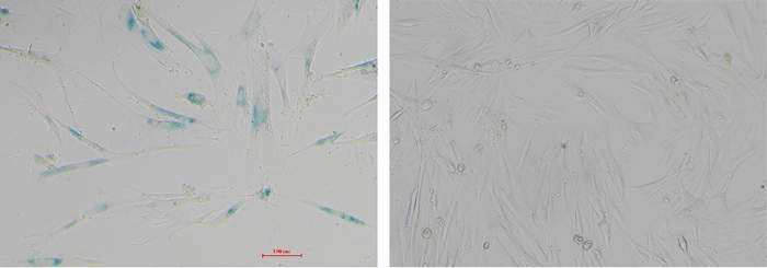
Fig.1 WI-38 cells were stained with β-galactosidase kit. The left picture shows senescence WI-38 cells without division and proliferation ability but still alive. After staining, the positive staining cells were more than 95%. The image on the right shows newly resuscitated WI-38 cells (early passage) with less than 3 passages, and no obvious positive cells after staining.
For Research Use Only!