No products in the cart.


15. Click-iT EdU-488 Cell Proliferation Assay Kit, 100 T(EdU-488) $650; 200T $900
$650.00
In Stock
Description
Product Introduction
Analyzing cell proliferation capability is a common and important assessment method in life sciences to determine the impact of genes, drugs, and other factors on cells cultured in vitro, or to analyze the growth and renewal ability of cells in biological tissues under different conditions or interventions. Currently, there are many methods available for detecting cell proliferation, most of which rely on the use of certain metabolic enzymes produced by cells to indirectly assess cell proliferation activity (such as the CCK-8 assay, MTT assay, etc.). However, factors such as drugs or the cellular state itself can influence the results of these assessments. Directly detecting DNA synthesis in cells to determine cell proliferation is recognized as the most accurate and effective detection method. However, both the originally used radioactive nucleoside incorporation method and the subsequent improved antibody-based BrdU method have their limitations.
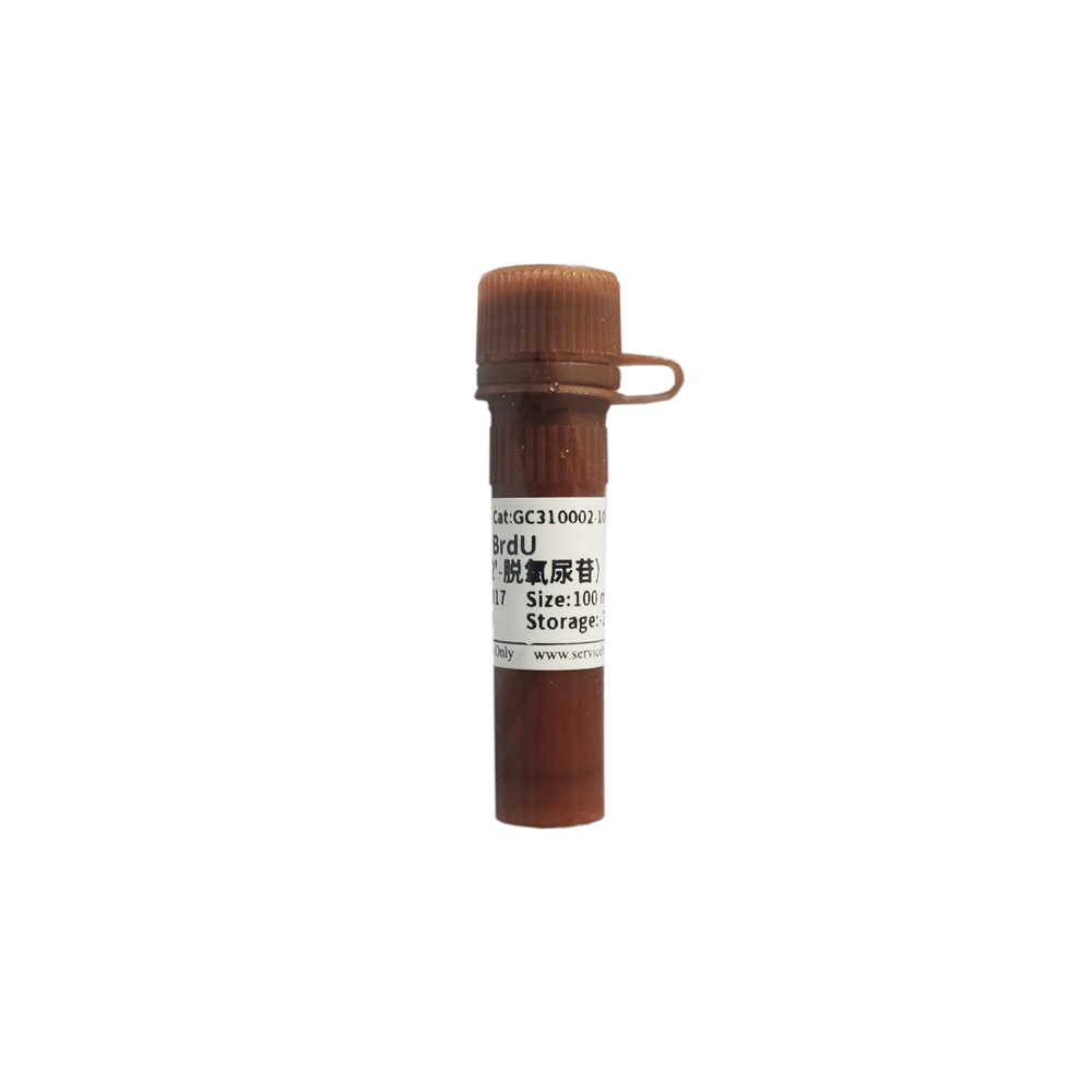
EdU (5-Ethynyl-2′-deoxyuridine) is a thymidine analog with an ethynyl group. When injected into animals or incubated with cells cultured in vitro, these small molecules can quickly diffuse into various organs and tissues and penetrate into cells. During cell proliferation, EdU can substitute for thymidine (T) and incorporate into newly synthesized DNA. The ethynyl group in the EdU molecule can undergo a “click” reaction with a fluorescent-labeled azide compound probe catalyzed by copper ions, forming a stable triazole ring, thereby allowing the newly synthesized DNA to be labeled by the corresponding fluorescent probe. Compared to the radioactive nucleoside incorporation method, the EdU detection method does not have limitations such as radioactive contamination. Compared to the BrdU detection method, the EdU detection method does not require DNA denaturation treatment or antigen-antibody reaction, significantly reducing the complexity and time required for the experiment, making it faster, more sensitive, stable, and accurate. This assay kit is used to detect cell proliferation in cells cultured in vitro or animal tissues. The fluorescent probe in this assay kit emits green fluorescence, with a maximum excitation wavelength of 491 nm and a maximum emission wavelength of 516 nm. After labeling proliferating cells, the cell nuclei will exhibit bright green fluorescence. The cell nuclei can be co-stained with a conventional nuclear dye (Hoechst 33342 provided in this assay kit), and the cell proliferation can be directly observed using fluorescence microscopy, laser confocal microscopy, or flow cytometry by detecting the fluorescence intensity of the cell population. The fluorescence intensity can be used to determine the DNA replication activity during the S phase of the cell cycle.
Storage and Transportation
Transportation with wet ice; store in the dark at -20°C. The EdU catalytic reagent (Reagent A) and reaction buffer can be stored at 4°C. Shelf life is 12 months.
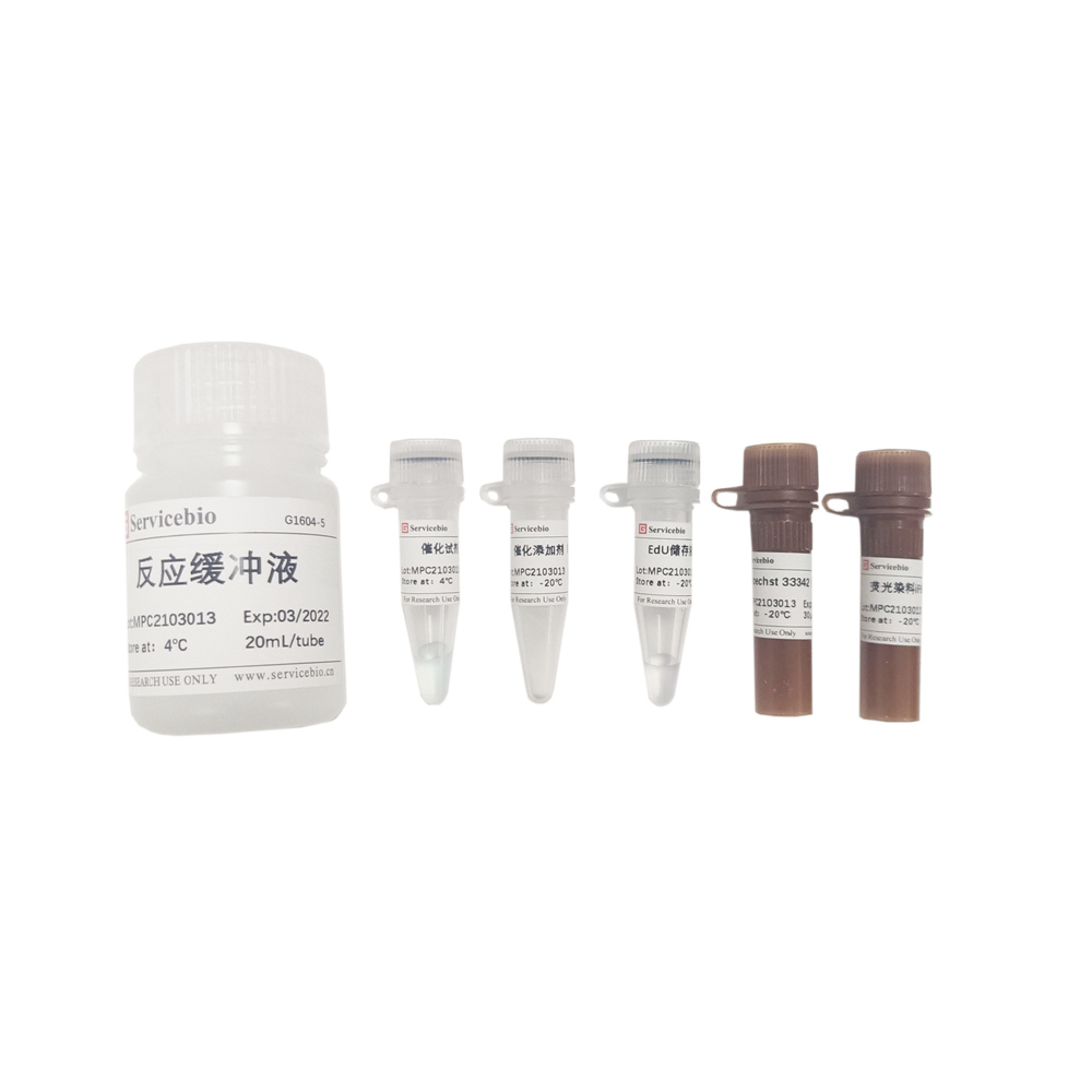
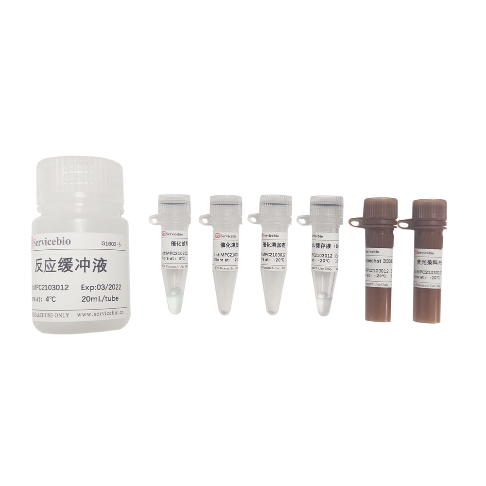
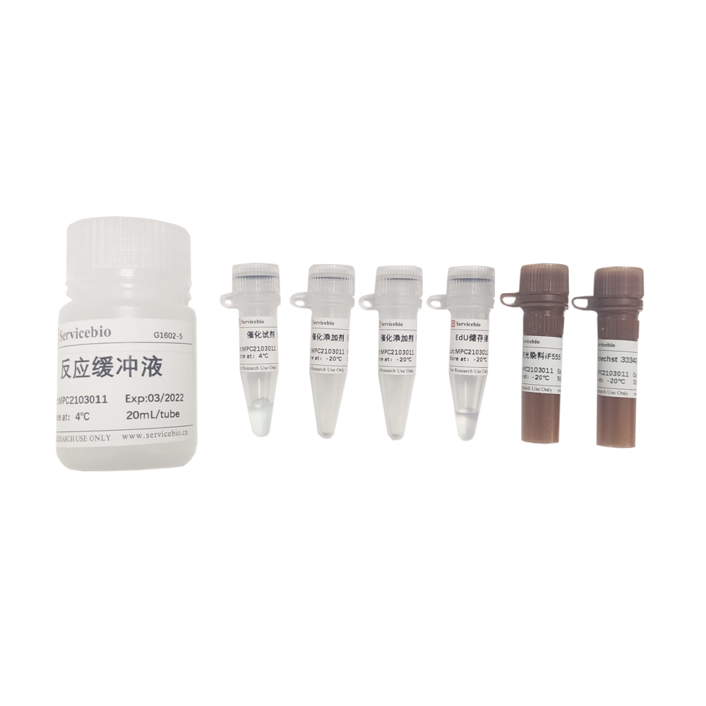
| Component Number | Component |
|---|---|
| G1601-100T | EdU Storage Solution (10 mM), 100 μL |
| G1601-1 | Catalytic Reagent (Reagent A), 120 μL |
| G1601-3 | Fluorescent Dye iF488 (Reagent B), 50 μL |
| G1601-4 | Catalytic Additive (Reagent C), 2×100 mg (powder) |
| G1601-5 | Reaction Buffer, 20 mL |
| G1601-6 | Hoechst 33342 Staining Solution, 30 μL |
| Product Manual | 1 copy |
Note: The above reaction numbers correspond to 96-well plate detection.
Experimental Preparation
- Serum-containing cell culture medium.
- Permeabilization solution: Buffer containing 0.2-0.5% Triton X-100 (recommended: G1204).
- Fixation solution: 4% paraformaldehyde (recommended: G1101) or similar reagent.
- PBS buffer (recommended: G4202).
- Ultrapure water.
- Reagents for animal modeling and tissue sectioning (for animal tissue cell proliferation detection).
Procedure
- Preparation of in vitro cell samples and reagents:
1.1. Seed cells evenly in cell culture plates at a certain density (the seeding density depends on factors such as cell size and growth rate). After the cells adhere or return to a normal state, perform the corresponding drug stimulation or other treatments (for suspended cells, follow the standard procedures for suspended cells, including centrifugation steps for the entire experiment).
1.2. Centrifuge the catalytic additive (reagent C) at low speed, take 100 mg, dissolve it in 1 mL ultrapure water, and store the aliquots at -20°C. The remaining powder can be kept as a backup.
- EdU labeling, fixation, and permeabilization of in vitro cells:
2.1. Prepare 2x EdU incubation working solution: Add 2 μL of EdU storage solution (10 mM) to 1 mL complete cell culture medium, resulting in a 20 μM 2x EdU incubation working solution. Preheat in a cell culture incubator (preliminary experiments are recommended using an EdU working concentration of 10 μM).
2.2. Using a partial medium exchange approach, remove half of the original cell culture medium from the culture plate and replace it with an equal volume of preheated 2x EdU incubation working solution. Incubate for a certain period of time (the incubation time generally depends on the cell’s growth cycle and is usually about 10% of the cell cycle duration. For most adherent cells with rapid growth, incubation for approximately 2 hours is recommended. Adjust the incubation time based on cell characteristics, treatment conditions, etc. If a longer incubation time is needed, decrease the EdU working concentration; for a shorter time, increase the EdU concentration accordingly).
2.3. After incubating the cells with EdU labeling, wash the samples 1-2 times with PBS buffer and add the fixation solution to cover the cells. Incubate at room temperature for 15 minutes (for flow cytometry analysis of adherent cells, perform digestion and resuspension before fixation, then follow the standard procedures for suspended cells). Wash 2-3 times with PBS buffer for 3-5 minutes each time.
2.4. Remove the PBS buffer, add the permeabilization solution to cover the cells or tissues, and incubate at room temperature for 15 minutes.
2.5. Remove the permeabilization solution, wash 1-2 times with PBS buffer for 3-5 minutes each time, then proceed to Step 4.
- Animal EdU Injection Modeling and Tissue Section Processing:
3.1. According to the experimental requirements, perform one or multiple EdU injections in animals through intraperitoneal, intramuscular, subcutaneous, or tail vein injection. The recommended ratio of EdU dosage to animal weight is 5 mg/kg, and the specific injection volume depends on the research content and the condition of the animal. This kit provides a portion of the EdU storage solution primarily for in vitro cell EdU labeling. If you need to perform EdU modeling in animals, you can separately purchase the EdU reagent (recommended: G5059).
3.2. Tissues with fast cell proliferation rates, such as the small intestine, and tissues with slow cell proliferation rates, such as the brain, have different labeling times. Generally, tissues with faster growth are labeled for less than 12 hours, while tissues with slower growth may require several days of labeling. The optimal labeling time depends on the specific experiment. Due to the fast proliferation of the small intestinal epithelial tissue, it is recommended to use such tissues as a reference for labeling time.
3.3. Sacrifice the animals according to the specified criteria, and then extract the desired tissues. Prepare frozen sections or paraffin sections following routine procedures.
a. For frozen sections: Allow the sections to reach room temperature, add an appropriate amount of fixation solution, and fix at room temperature for 15 minutes. Remove the fixation solution, wash with an appropriate amount of PBS buffer for 3-5 minutes, three times. Remove the PBS buffer, cover the tissue with an appropriate amount of permeabilization solution, and incubate at room temperature for 10-15 minutes. Remove the permeabilization solution, wash with PBS buffer for 3-5 minutes, one or two times, and then proceed to Step 4.
b. For paraffin sections: Deparaffinize and rehydrate the sections, wash with PBS for 5 minutes. Remove the PBS buffer, add the permeabilization solution to cover the cells or tissues, and incubate at room temperature for 15 minutes. After removing the permeabilization solution, wash with PBS buffer for 3-5 minutes, one or two times, and then proceed to Step 4.
- EdU Click Reaction:
4.1. During the fixation and permeabilization of cells or tissues, prepare the click reaction mixture according to the following table:
For in vitro cultured cells: The reference step is based on the volume for 10 samples in a 96-well plate (100 μL per well). Adjust the amounts proportionally based on your needs. Add the components in the order listed below while gently mixing (prepare and use immediately).
| Component | Volume |
|---|---|
| Reaction Buffer | 935 μL |
| Catalyst (Reagent A) | 10 μL |
| Fluorescent Dye iF488 | 5 μL |
| Catalyst Additive (Reagent C) | 50 μL |
| Total Volume | 1000 μL |
For tissue cell sections, refer to the following system for the preparation of the click reaction solution. The preparation volume can be proportionally increased or decreased based on the number of tissue samples, with approximately 100-200 μL of click reaction solution covering each section.
| Component | Volume |
|---|---|
| Reaction Buffer | 928 μL |
| Catalyst (Reagent A) | 10 μL |
| Fluorescent Dye iF488 (Reagent B) | 12 μL |
| Catalyst Additive (Reagent C) | 50 μL |
| Total Volume | 1000 μL |
4.2. Remove the previous PBS buffer (from step 2.5 or 3.3), add the Click reaction mixture, gently shake to ensure that the reaction mixture covers all cells or tissues, and incubate at room temperature in the dark for 30 minutes.
4.3. Remove the Click reaction mixture, wash with PBS buffer 2-3 times, for 3-5 minutes each time (if there are no specific requirements, the fluorescence intensity can be detected using flow cytometry or other instruments).
- Nuclear staining:
5.1. Remove the previous PBS buffer, dilute Hoechst 33342 staining solution with PBS buffer at a ratio of 1:500-1000, add it to cover the cells, and incubate for 5 minutes.
5.2. Remove the Hoechst 33342 staining solution, wash with PBS buffer 2-3 times, for 3-5 minutes each time.
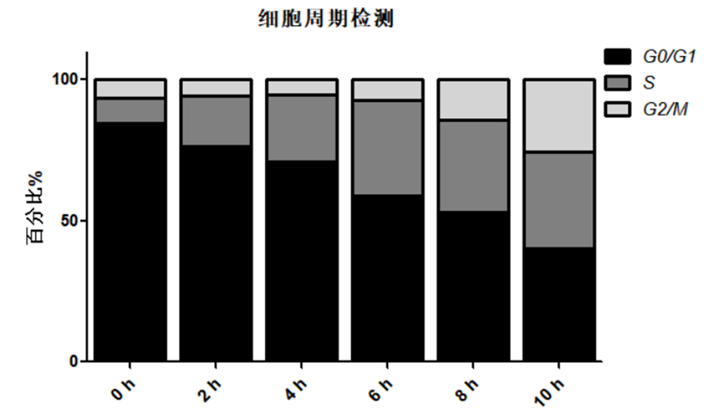
- Imaging and Detection Analysis
Directly place the cultured cells or tissue slice samples in a fluorescence microscope or confocal microscope for detection, and analyze the proportion of proliferating cells. Alternatively, collect the cultured cells and detect the fluorescence intensity using flow cytometry (it is recommended to use non-labeled cells as a negative control for flow cytometry detection, and select appropriate voltage settings). Based on the fluorescence intensity, the DNA replication activity during the S phase of the cell cycle can be determined. The fluorescent dye iF488 (Reagent B) in this kit has a spectral characteristic of Ex/Em: 491 nm/516 nm (green), and Hoechst 33342 staining solution has a spectral characteristic of Ex/Em: 346 nm/460 nm (blue).
Notes:
- For cultured cells, the specific concentration of EdU and incubation time may vary depending on the sample and research purpose and can be adjusted accordingly.
- Some tissue cells have slow proliferation rates, so to exclude factors such as poor modeling effects, it is recommended to select tissue samples with fast proliferation rates as reference samples (such as intestinal tissue).
- If the background color is too dark, it may be due to insufficient washing during the experiment, excessive fixation time of tissue samples, or residual fixative.
- The catalyst additive (Reagent C) of EdU is prone to oxidation, so it is recommended to avoid prolonged exposure to air. After preparing it into an aqueous solution, it is advisable to store it in aliquots. Based on tests, if the color of the catalyst additive of EdU undergoes slight changes, the Click reaction catalytic system can still proceed normally. However, if it turns brown, it indicates that the component has become ineffective and should be discarded.
- Please wear lab coat and disposable gloves while performing the procedure.
This product is for research use only and is not intended for clinical diagnosis.
Version: V1.0-202103