No products in the cart.


14. Cell Cycle and Apoptosis Detection Kit, 50 T $249
$249.00
In Stock
Description
Product IntroductionRelated Products
Product Information
| Product Name | Cat.No | Spec. |
| Cell Cycle and Apoptosis Detection Kit | G1700-50T | 50 T |
Introduction
Cell cycle refers to the whole process of the cell from the completion of one division to the end of the next division, which is mainly divided into two stages: intermitotic phase and mitotic phase (M phase). The intercellular phase is mainly composed of the early phase of DNA synthesis (G1 phase), the late phase of DNA synthesis (S phase) and the late phase of DNA synthesis (G2 phase). The sequence of the whole cell cycle can be expressed as G1→S→G2→M. First of all, in G1 phase, cells mainly synthesize RNA and proteins to prepare materials and energy for cells to enter S phase. Then it enters S phase: the cells begin to synthesize DNA and some histones and other substances, and the cell DNA content begins to increase. Finally, the G2 phase: at this time, the DNA content of the cell has become twice that of the G1 phase, and has stopped DNA replication, to enter the mitotic phase to do a lot of protein and other material synthesis; If the cell is in the G0(Cells temporarily stop dividing, differentiating, and quiescence)/G1 phase, the DNA content of the cell is 1N; So cells in G2 have 2N DNA; The DNA content of S-phase cells between G1 and G2 was between 1N and 2N. In apoptotic cells, the nucleus will be concentrated and DNA fragmentation will occur, leading to the loss of part of genomic DNA fragments, so the DNA content is less than 1N, and the so-called sub-G1 peak, namely the apoptotic cell peak, appears on the fluorescence map detected by flow cytometry. Therefore, the cell cycle and state can be judged according to the content of cell DNA.
Apoptosis can also be detected by observing changes in cell light scattering by flow cytometry. When apoptosis occurs, the cytoplasm and chromatin are concentrated and the nucleus is fragmented, resulting in apoptotic bodies. Chromatin shrinks and cell density increases in pre-apoptosis stage. In the late stage of apoptosis, cells produce apoptotic bodies, and the light scattering of cells changes.
The Cell Cycle and Apoptosis Analysis Kit used the classic Propidium staining method to detect and analyze Cell Cycle and Apoptosis. Propidium iodide is able to embed in double-stranded DNA and cause it to fluoresce. The cycle and state of a cell can be distinguished by the characteristic that the fluorescence intensity is proportional to the content of double-stranded DNA and the regular change of DNA content in different cell cycles. This kit can be used for cell cycle and apoptosis detection of tissue cells, adherent or suspended cells (if used for cell cycle and apoptosis detection of tissue, the tissue must be digested into a single cell state before detection).
Storage and Handling Conditions
Transport with wet ice ; Stored at -20℃ away from light, dyeing buffer can be stored at 4℃; It is valid for 12 months.
Components
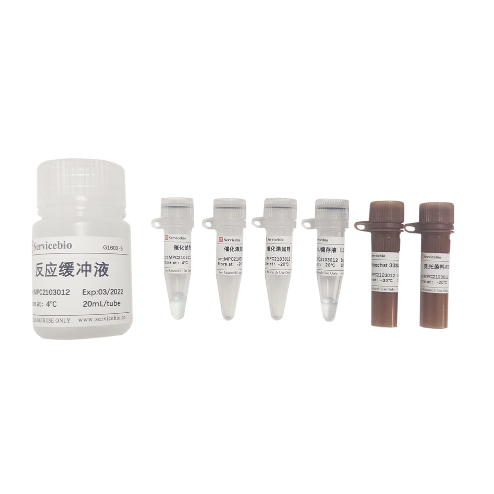
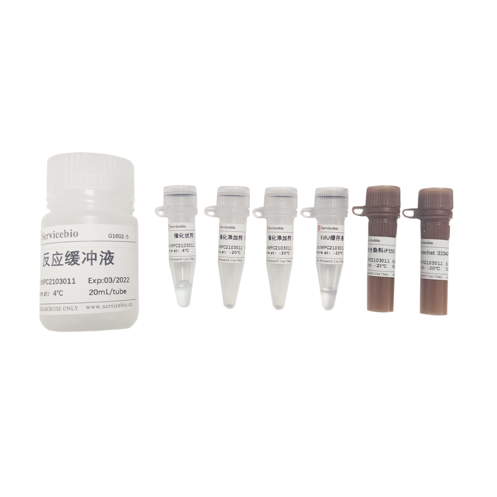
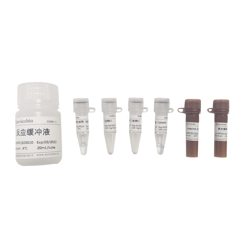
| Component Number | Component | G1700-50T |
| G1700-1 | PI Staining Solution (50×) | 500 μL |
| G1700-2 | RNase A (50×) | 500 μL |
| G1700-3 | Staining Buffer | 25 mL |
| Instruction Manual | 1 pc | |
Notice Before Using (Please Read Carefully)
1. Cell culture medium containing serum;
2. Trypsin digestion solution (G4001 was recommended);
3. PBS buffer (G4202 was recommended);
4. 75% ethanol.
Assay Protocol / Procedures
1. Preparation of Cell Samples (the number of cells is controlled at 1×10⁵~1 ×10⁶)
1.1. For adherent cells: remove the culture medium, add trypsin digestion solution to digest the cells, and observe that the cells become round and loose under the microscope. The digestion was terminated by adding proper amount of cell medium containing serum, and the cells were gently blown away to make the suspension. The suspension was transferred to a centrifuge tube, centrifuged at 1000 g for 3-5 min, and the supernatant was discarded to retain the cell precipitation. Then the cell precipitation was washed 1-2 times with pre-cooled PBS buffer, and the supernatant was discarded by centrifugation in the same way to retain the cell precipitation.
1.2. For the suspended cells: transfer the cells to a centrifuge tube, centrifuge at 1000 g for 3-5 min, discard the supernatant, and retain the cell precipitate; Then the cell precipitation was washed 1-2 times with pre-cooled PBS buffer, and the supernatant was discarded by centrifugation in the same way to retain the cell precipitation.
1.3. For tissue cells: Cut the tissue into as small pieces as possible. According to the source of tissue, the tissue block was digested by trypsin, collagenase and other digestive enzymes, and the single cell suspension was obtained by filtering through 100-300 mesh screen. The filtered cell suspension was transferred to a centrifuge tube, centrifuged at 1000 g for 3-5 min, and the supernatant was discarded to retain the cell precipitation. Then the cell precipitation was washed 1-2 times with pre-cooled PBS buffer, and the supernatant was discarded by centrifugation in the same way to retain the cell precipitation.
2. Fixation of Cell Samples
2.1. Add 1 mL of 75% ethanol pre-cooled on ice to the collected cell precipitation sample, and gently blow the cells to make them thoroughly mixed;
2.2. The cells were fixed at 4℃ for 30 min or longer (usually 2 h or more can guarantee the staining effect, and 12-24 h may be better to improve the staining effect).
2.3. After fixed for a certain time, the cells were centrifuged at 1000 g for 3-5 min to remove the ethanol fixing solution and retain the cell precipitate;
2.4. Tap the bottom of the centrifuge tube to disperse the cells, resuspended and washed the cells with PBS buffer, centrifuged at 1000 g for 3-5 min, and discarded the supernatant to collect cell precipitation;
3. Preparation and Staining of Working Solution
3.1. According to the following table, the dyeing solution can be prepared out of light, and the amount can be increased or decreased in equal proportion according to the use requirements (the prepared dyeing solution can be stored at 4℃ in a short time, please use it within the same day);
| 1 Sample | 5 Samples | 10 Samples | |
| Staining Buffer | 480 μL | 2.4 mL | 4.8 mL |
| PI Stain (50×) | 10 μL | 50 μL | 100 μL |
| RNaseA (50×) | 10 μL | 50 μL | 100 μL |
| Total Volume | 500 μL | 2.5 mL | 5 mL |
3.2. Tap the bottom of the centrifuge tube to disperse the cells precipitated in Step 2.4, then add 500 μL of the prepared staining working solution, and gently blow to disperse the cells and mix with the staining working solution;3.3. After incubation at 37℃ for 30 min in the dark, flow cytometry can be used for detection.
4. Flow Detection and Analysis
The red fluorescence was detected by flow cytometry at the excitation wavelength of 488nm, and the light scattering was also detected. Cell DNA content analysis and light scattering analysis were performed using appropriate analysis software.
Note
1. All fluorescent dyes have the problem of fluorescence quenching, so try to avoid light during use and storage.
2. Before the experiment, it is suggested to synchronize the cell cycle to avoid large reproducibility differences caused by different cell cycles.
3. The planting density of experimental cells should not be too high or too low to prevent contact inhibition or density dependence.
4. Pay attention to protection when operating PI staining solution, avoid direct contact with human body or inhalation.
5. For your safety and health, please wear lab coat and disposable gloves when operating.
For Research Use Only!
Product recommendation
|
Cat.No.
|
Product Name
|
Spec.
|
Operation
|
|---|
|
G1501-100T
|
Fluorescein (FITC) Tunel Cell Apoptosis Detection Kit
|
100 T
|
|
|
G1501-50T
|
Fluorescein (FITC) Tunel Cell Apoptosis Detection Kit
|
50 T
|
|
|
G1502-100T
|
TMR (Red) Tunel Cell Apoptosis Detection Kit
|
100 T
|
|
|
G1502-50T
|
TMR (Red) Tunel Cell Apoptosis Detection Kit
|
50 T
|
|
|
G1504-100T
|
CF488 Tunel Cell Apoptosis Detection Kit
|
100 T
|
|
|
G1504-50T
|
CF488 Tunel Cell Apoptosis Detection Kit
|
50 T
|
|
|
G1505-100T
|
CF640 Tunel Cell Apoptosis Detection Kit
|
100 T
|
|
|
G1505-50T
|
CF640 Tunel Cell Apoptosis Detection Kit
|
50 T
|
|
|
G1507-100T
|
DAB (SA-HRP) Tunel Cell Apoptosis Detection Kit
|
100 T
|
|
|
G1507-50T
|
DAB (SA-HRP) Tunel Cell Apoptosis Detection Kit
|
50 T
|
|
|
G1510-100T
|
Annexin V-EGFP/PI Cell Apoptosis Detection Kit
|
100 T
|
|
|
G1510-50T
|
Annexin V-EGFP/PI Cell Apoptosis Detection Kit
|
50 T
|
|
|
G1511-100T
|
Annexin V-FITC/PI Cell Apoptosis Detection Kit
|
100 T
|
|
|
G1511-50T
|
Annexin V-FITC/PI Cell Apoptosis Detection Kit
|
50 T
|
|
|
G1512-100T
|
Annexin V-PE/7-AAD Cell Apoptosis Detection Kit
|
100 T
|
|
|
G1512-50T
|
Annexin V-PE/7-AAD Cell Apoptosis Detection Kit
|
50 T
|
|
|
G1513-100T
|
Annexin V-IF488/PI Cell Apoptosis Detection Kit
|
100 T
|
|
|
G1513-50T
|
Annexin V-IF488/PI Cell Apoptosis Detection Kit
|
50 T
|
|
|
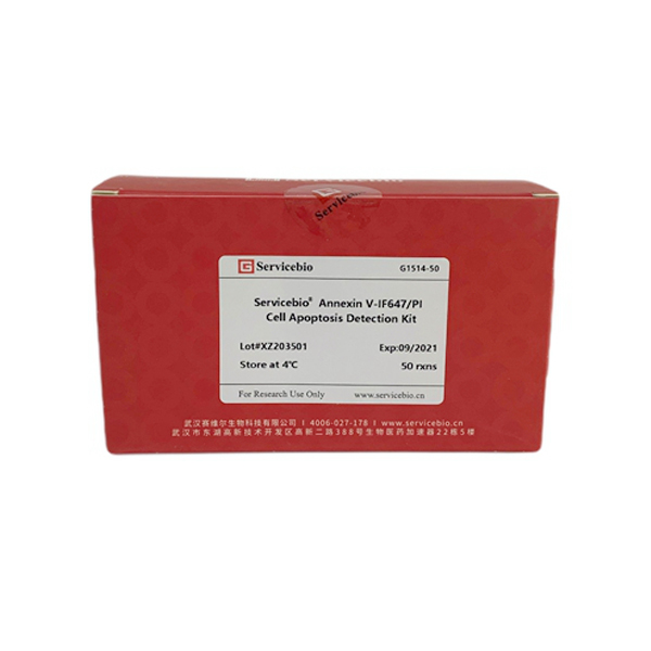
G1514-100T
|
Annexin V-IF647/PI Cell Apoptosis Detection Kit
|
100 T
|
|
|
G1514-50T
|
Annexin V-IF647/PI Cell Apoptosis Detection Kit
|
50 T
|
|
|
G1700-50T
|
Cell Cycle and Apoptosis Detection Kit
|
50 T
|