No products in the cart.

15. Fluorescent Lipid Droplet Assay Kit, $599
$599.00
In Stock
Description
Fluorescent Lipid Droplet Detection Assay Kit
Product Code: G1905-100T
Specification: 100 tests
Product Description:
Lipid droplets are specialized organelles within cells that store lipids, regulating the storage and hydrolysis of neutral lipids to provide dynamic energy balance for cells. Additionally, lipid droplets can move along the cell’s cytoskeleton, interact with other organelles, and play a significant role in signal transduction regulation. Abnormal accumulation of cellular lipid droplets occurs in various metabolic disorders such as obesity, fatty liver, cardiovascular diseases, and pathological states.
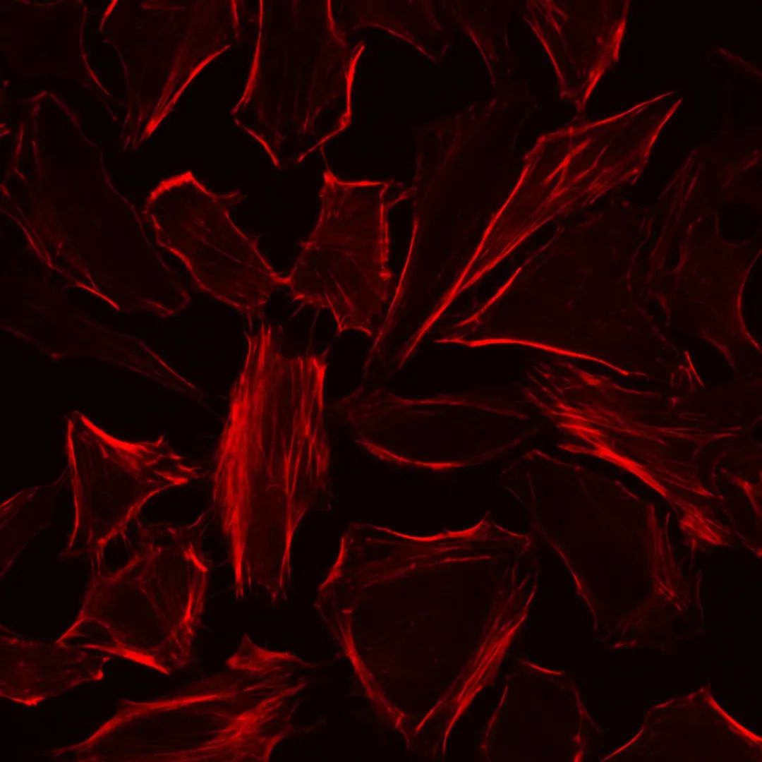
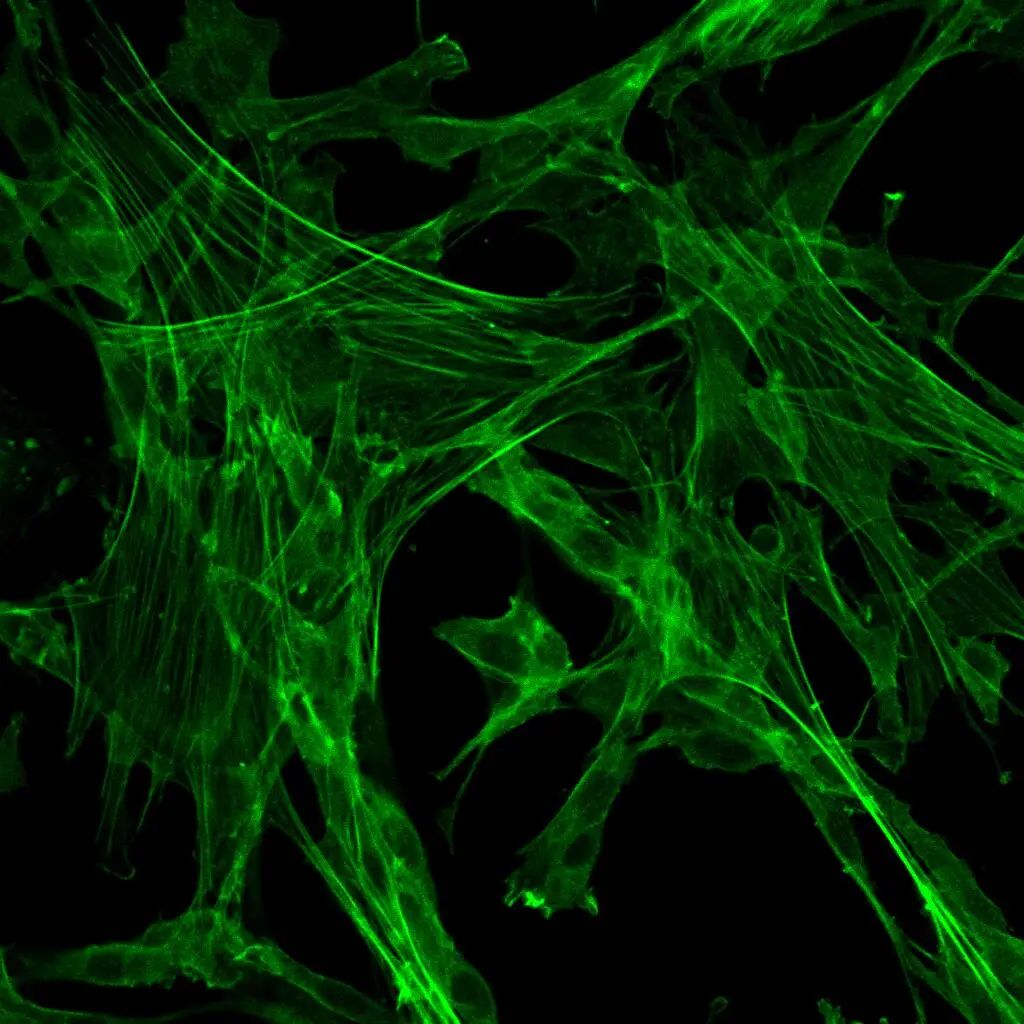
The Fluorescent Lipid Droplet Detection Assay Kit utilizes BODIPY 493/503 fluorescent probe as its core component. This probe is a lipophilic fluorescent dye that localizes to polar lipids, making it suitable for quantifying neutral lipid content in cells and tissues. Developed and optimized by our research team, this assay kit offers advantages such as ease of use, stability, and good reproducibility. The higher the cellular lipid content, the stronger the fluorescence intensity generated. Detection can be performed using instruments such as fluorescent microscopes or flow cytometers. Additionally, the kit provides oleic acid as an inducer for triglyceride synthesis and storage, serving as a positive control inducer.
Storage and Transportation:
Shipment on dry ice; store at -20°C, avoid light exposure, shelf life of 6 months.
Kit Components:
Component Number
Component
G1905-100T
G1905-1
Lipid Droplet Detection Probe (2 mM)
100 μL
G1905-2
Lipid Droplet Detection Buffer
100 mL
G1905-3
Positive Control Inducer (500 mM)
50 μL
Note: The above reaction amounts are for a 6-well plate detection system.
Operational Steps:
Note: The following steps are given for adherent cells in a 6-well plate. For suspension cells, centrifugation steps are required during media changes. For tissue samples, refer to the staining and labeling steps, and adjust overall reagent volumes accordingly. If necessary, you can fix the samples in advance, but avoid perforating them.
- Preparation of Lipid Droplet Detection and Induction Working Solutions:
1.1. Mix an appropriate volume of Lipid Droplet Detection Probe (2 mM) with Lipid Droplet Detection Buffer in a 1:500-2000 ratio to prepare the Lipid Droplet Detection working solution.
1.2. Dilute an appropriate volume of Positive Control Inducer (500 mM) with the corresponding cell culture medium to achieve a concentration of 400-1000 μM, preparing the Lipid Droplet Induction working solution (this concentration is a reference range and can be adjusted based on cell properties or other factors).
- Positive Control and Treatment Groups (given as an example for a 6-well plate, adjust steps accordingly for suspension cells):
2.1. Prepare cells in a 6-well plate as needed (consider subculturing or not based on detection method and equipment).
2.2. Remove original cell culture medium, wash twice with PBS or other buffer.
2.2.1. Experimental Group: Stimulate test cells with drugs or other relevant treatments and incubate as required.
2.2.2. Positive Control Group: Add 1 mL of Lipid Droplet Induction working solution to each well and incubate overnight.
2.3. After incubation, proceed to Step 3 for staining and labeling.
- Lipid Droplet Staining and Labeling:
3.1. Remove cells’ original medium, wash 2-3 times with PBS or other buffer.
3.2. Remove washing buffer, add 1 mL of Lipid Droplet Detection working solution, mix gently.
3.3. Incubate in the dark at room temperature or a CO2 incubator for 15 minutes.
3.4. After incubation, wash 2 times with PBS or other buffer. Proceed to further staining or analysis using appropriate equipment.
- Lipid Droplet Detection:
4.1. Analyze the incubated and washed samples using the relevant instrument, observing green fluorescence after labeling. Example instrument detection methods include:
4.1.1. Directly observe using a fluorescent microscope or perform analysis after sealing with anti-fade mounting medium (Catalog No.: G1401).
4.1.2. Perform cell digestion with trypsin solution (for adherent cells) or directly analyze (for suspension cells) using a flow cytometer.
Precautions:
- Due to variations in cell types and properties, adjust the concentration of positive control inducer and probe, as well as the incubation time, based on experimental needs.
- Prevent sample contact with organic solvents or other components that might affect lipid substances to maintain accurate detection.
- Be cautious of fluorescence quenching during the operation process; avoid light exposure.
- This product is for research purposes only and not intended for clinical diagnosis or treatment.
This product is for research use only, not for clinical diagnosis.