No products in the cart.

13. MitoTracker Red CMXRos (Mitochondrial Red Fluorescent Probe), $599
$599.00
In Stock
Description
Product Information
Product Name: MitoTracker Red CMXRos (Mitochondrial Red Fluorescent Probe) Product Number: G1723-50UG Specification: 50 μg
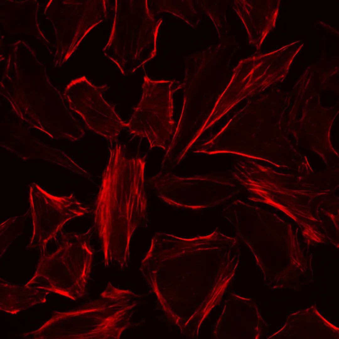
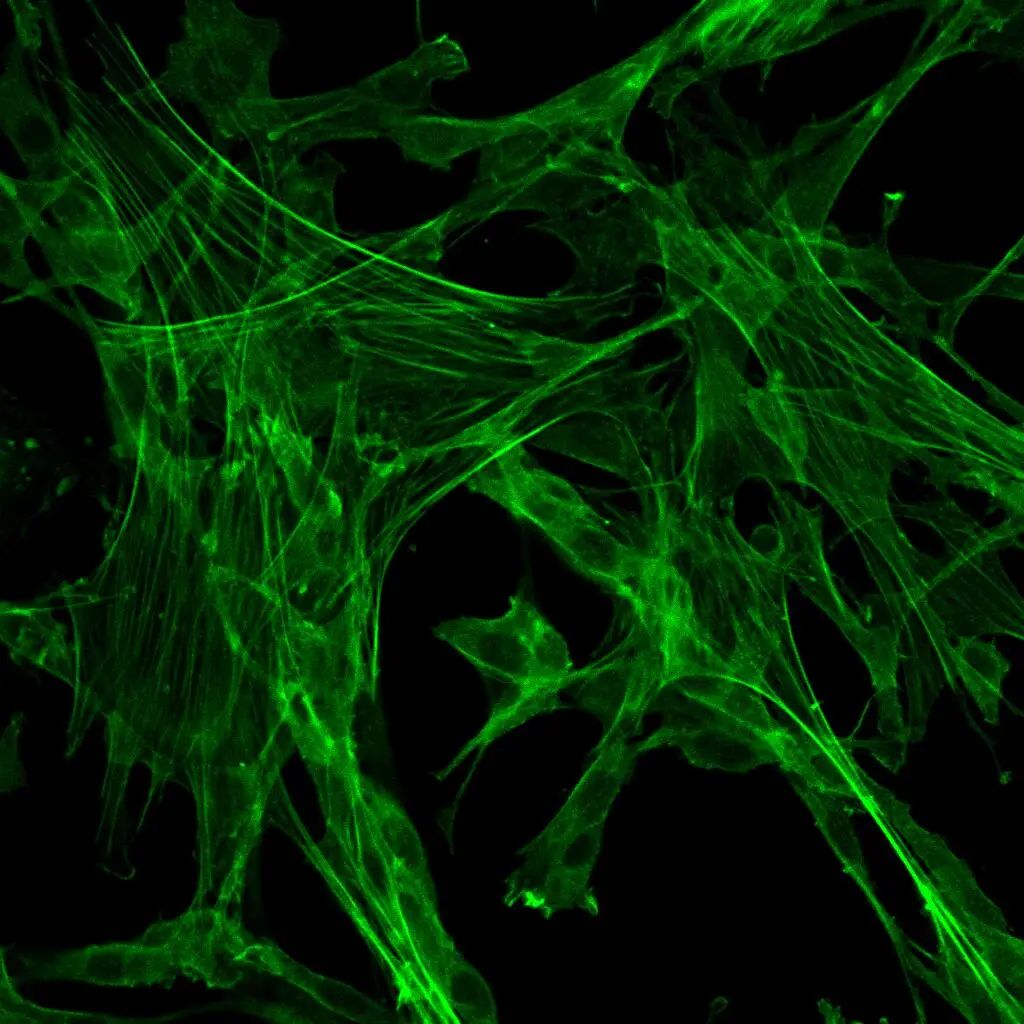
Product Description: MitoTracker Red CMXRos is a derivative of X-rosamine with a molecular weight of 531.52. It has an excitation wavelength of 578±3 nm and an emission wavelength of 599±4 nm. This dye can directly enter live cells and localize to mitochondria, where it covalently binds to the mitochondria. The specific labeling principle of mitochondria is based on the positively charged probe backbone, which allows the probe to target the negatively charged mitochondria. The chloromethyl group on the probe can covalently bind to free thiol groups within the mitochondria, resulting in fluorescent labeling. Simply incubating the probe with live cells for a certain period of time can label cellular mitochondria. After mitochondrial labeling, the cells can undergo fixation and permeabilization for further multi-label experiments.
Storage and Transportation: Shipped on wet ice; store at -20°C in the dark. The product is valid for 12 months.
Kit Components:
| Component | Quantity |
|---|---|
| MitoTracker Red CMXRos (线粒体红色荧光探针) | 50 μg |
| Product Manual | 1 copy |
Operational Steps:
- Preparation of Detection Working Solution:
- Note: This product is in powder form and needs to be briefly centrifuged at high speed before use to ensure that the powder settles at the bottom of the tube. Prepare cell-grade DMSO.
- Prepare storage solution: Add 94 μL of cell-grade DMSO into the tube, mix by pipetting to ensure complete dissolution of the dye, resulting in a 1 mM concentration of the mitochondrial fluorescent dye storage solution (molecular weight information is provided on the product label). Centrifuge briefly to prevent reagent loss due to attachment to the walls. Store the solution at -20°C. It is recommended to aliquot into smaller volumes to avoid repeated freeze-thaw cycles.
- Prepare detection working solution: If the staining will be directly followed by detection, dilute the storage solution with biological buffer (Hanks, PBS) or serum-free basal medium to obtain a final concentration of 0.1-0.25 μM of the mitochondrial detection working solution. If subsequent fixation and permeabilization are required after staining, dilute the storage solution to a final concentration of 0.4-0.6 μM of the mitochondrial detection working solution.
- Cell Staining (below is for adherent cells; centrifugation is required for suspension cells):
- Seed cells on cell culture plates or coverslips until they reach a suitable density.
- Aspirate the cell culture medium and wash the cells 1-2 times with pre-warmed buffer, 3-5 minutes per wash.
- Add pre-warmed mitochondrial detection working solution, incubate at 37°C for 30 minutes (adjust incubation time based on cell type and condition).
- Remove the incubation solution and wash the cells 2-3 times with buffer, 3-5 minutes per wash.
- Observe the cells using fluorescence microscopy directly or after sealing with anti-fluorescence quenching sealing solution (recommended G1401) (Ex=579 nm, Em=599 nm).
- Cell Fixation, Permeabilization, and Other Operations (optional):
- Note: After staining, cells can undergo fixation, permeabilization, and other operations, but fluorescence signal may attenuate after fixation.
- Cell Fixation:
- Add fixed solution (recommended G1101) to cells stained and washed with mitochondrial fluorescent dye, fix at room temperature for 10 minutes.
- Remove the fixed solution and wash the cells 2-3 times with buffer, 3-5 minutes per wash.
- Cell Permeabilization:
- Add 0.2-0.5% Triton X-100 to fixed and washed cells, permeabilize at room temperature for 10 minutes.
- Remove the permeabilization solution and wash the cells 2-3 times with buffer, 3-5 minutes per wash.
- After staining, fixing, and permeabilization, cells can be used for subsequent multi-label experiments.
Important Notes:
- Due to variations in cell types and states, the dye concentration and incubation time can be adjusted accordingly.
- This dye requires live-cell status for mitochondrial localization; cells should not be fixed prior to staining to avoid false-positive results caused by inaccurate localization. Fixation and subsequent operations can be performed after mitochondrial staining.
- When staining and washing live cells, pre-warm the detection working solution and washing buffer to 37°C. Staining and washing should be completed in an incubator to avoid temperature fluctuations that might affect cell morphology.
- To prevent fluorescence quenching, the entire staining process should be performed in the absence of light.
- Probe stock solution should be aliquoted and stored to avoid repeated freeze-thaw cycles. Staining working solution should be prepared as needed.
- For your health and safety, wear appropriate laboratory attire and gloves while working with this product.
- This product is intended for research purposes only and is not for clinical diagnosis.
(Note: Product packaging is being upgraded; refer to the actual product for details.)