Description
Product Information
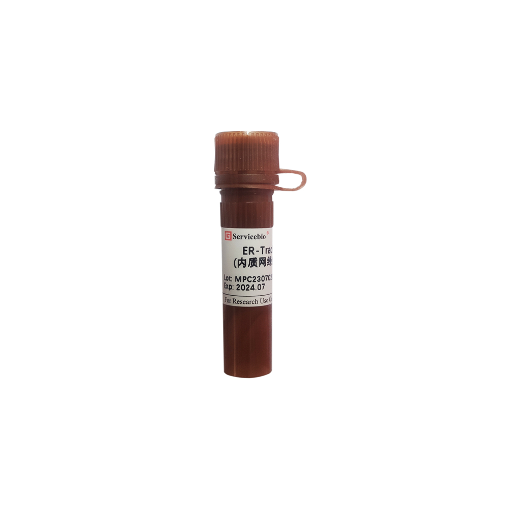
Product Name: ER-Tracker Green (Endoplasmic Reticulum Green Fluorescent Probe) Product Number: G1720-20UL Specification: 20 μL
Product Description: ER-Tracker Green is a fluorescent probe that is composed of the green fluorescent dye BODIPY FL and Glibenclamide. The chemical structure includes the Glibenclamide moiety, originally used as a type II diabetes treatment drug, which can bind to sulfonylurea receptors on the ATP-sensitive potassium ion channels of the endoplasmic reticulum. This allows the probe to selectively localize to the endoplasmic reticulum, serving as a specific fluorescent probe for labeling the endoplasmic reticulum in cells. ER-Tracker Green exhibits good specificity, particularly for the endoplasmic reticulum in live cells, with minimal mitochondrial staining and low cytotoxicity. After staining live cells, this probe can partially preserve the staining characteristics of viable cells.
Storage and Transportation: Shipped on dry ice; Store at -20°C in a light-protected environment; Shelf life of 12 months.
Kit Components:
Component:
G1720-20UL ER-Tracker Green (Endoplasmic Reticulum Green Fluorescent Probe) 20 μL
Product Manual: 1 copy
Operating Steps:
- Preparation of Detection Working Solution:
1.1 This product is stored as a 1 mM solution. Before use, warm to room temperature, briefly centrifuge at low speed to ensure the reagent settles at the bottom of the tube.
1.2 Dilute the reagent with buffer (such as Hanks’ balanced salt solution or PBS, with Hanks’ recommended) or basic culture medium (serum-free) to a concentration of 0.25-1 μM for the endoplasmic reticulum detection working solution.
- Cell Staining (the following protocol is for adherent cells; for suspension cells, centrifugation steps are needed):
2.1 Wash normal or treated cells with buffer 1-2 times, each time for 3-5 minutes.
2.2 Add pre-warmed endoplasmic reticulum detection working solution and incubate in a cell culture incubator at 37°C for 20 minutes (adjust the incubation time according to cell type and condition).
2.3 Remove the endoplasmic reticulum detection working solution and wash cells with buffer 2-3 times, each time for 3-5 minutes.
2.4 Observe green fluorescence (Ex=504 nm, Em=511 nm) directly or after sealing with anti-fluorescence quenching reagent (recommended G1401) using a fluorescence microscope.
- Cell Fixation (optional):
Note: After staining, the cells can be fixed; however, fixation may lead to attenuation of fluorescence signal. Permeabilization should be avoided as it leads to loss of fluorescence labeling.
3.1 Add cells after staining and washing to fixative solution (recommended G1101) and fix at room temperature for 5 minutes.
3.2 Remove fixative solution and wash cells with buffer 2-3 times, each time for 3-5 minutes.
3.3 Process fixed cells as needed for subsequent microscopy, slide sealing, or further staining.
Precautions:
- Due to variations in cell types and conditions, the working concentration of the dye and incubation time can be adjusted accordingly.
- This probe cannot be used for permeabilization after staining, as permeabilization would almost completely eliminate the cell fluorescence.
- The pharmacological properties of Glibenclamide may affect certain functions of the endoplasmic reticulum. Variable expression of sulfonylurea receptors in specific cells may lead to non-endoplasmic reticulum-specific staining.
- Warm the detection working solution and washing buffer to 37°C before staining and washing. Staining and washing should be conducted in a cell culture incubator to avoid temperature shock affecting cell morphology.
- To prevent fluorescence quenching, the entire staining process should be carried out in the absence of light.
- The probe solution should be aliquoted to avoid repeated freeze-thaw cycles. The staining working solution should be prepared as needed and used immediately.
- For your health and safety, please wear appropriate lab attire and gloves during operation.
This product is for research purposes only and is not intended for clinical diagnosis.
(Product packaging is being upgraded, actual product may differ.)

