No products in the cart.


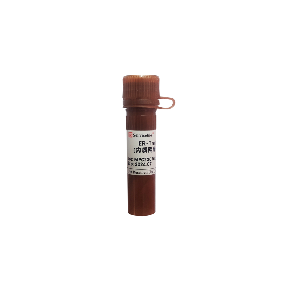
12. ER-Tracker Red (Endoplasmic Reticulum Red Fluorescent Probe), $599
$599.00
In Stock
Description
Product Information
Product Name: ER-Tracker Red (Endoplasmic Reticulum Red Fluorescent Probe) Product Number: G1721-20UL Specifications: 20 μL
Product Description:
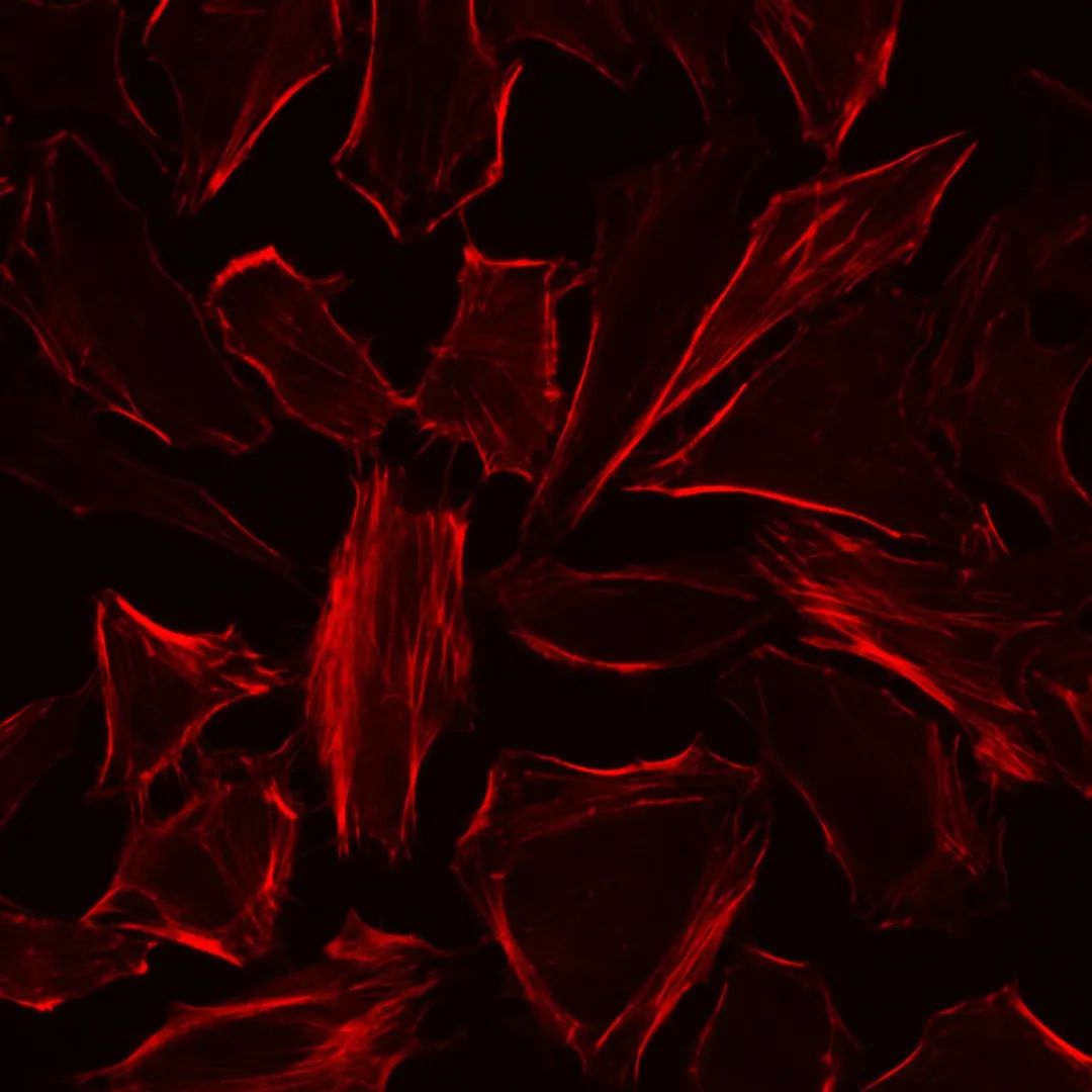
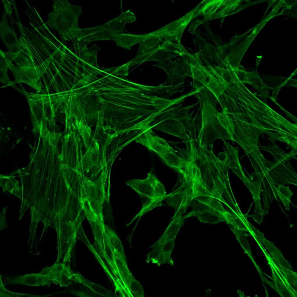
ER-Tracker Red is a red fluorescent probe that is used for specific staining of the endoplasmic reticulum (ER) in cells. This probe consists of a red fluorescent dye BODIPY TR and Glibenclamide, a sulfonylurea receptor known for its binding to ATP-sensitive potassium channels on the ER membrane. This property allows the probe to selectively target the ER, making it a specific tool for labeling the endoplasmic reticulum. ER-Tracker Red exhibits good specificity for the ER in live cells, with minimal staining of mitochondria and low cell toxicity. The probe can retain some staining characteristics of live cells after fixation.
Storage and Transport:
ER-Tracker Red is typically shipped on dry ice and should be stored at -20°C, protected from light, to maintain its stability and effectiveness for up to 12 months.
Components of the Kit:
- G1721-20UL: ER-Tracker Red (Endoplasmic Reticulum Red Fluorescent Probe) – 20 μL
- Product Manual: 1 copy
Operating Steps:
- Preparation of Detection Working Solution:
1.1 The product is provided as a 1 mM stock solution. Before use, thaw the product to room temperature, briefly centrifuge it at low speed to ensure that the reagent is at the bottom of the tube. 1.2 Dilute the stock solution with a suitable buffer (such as Hanks or PBS) or basic culture medium (serum-free) to obtain a 0.25-1 μM ER detection working solution.
- Cell Staining (Procedure for adherent cells; for suspension cells, centrifugation steps are required):
2.1 Wash normal or treated cells with buffer solution 1-2 times, each time for 3-5 minutes. 2.2 Add pre-warmed ER detection working solution and incubate at 37°C for 20 minutes (adjust incubation time as needed based on cell type and condition). 2.3 Remove the ER detection working solution and wash cells with buffer solution 2-3 times, each time for 3-5 minutes. 2.4 Observe red fluorescence (Ex=587 nm, Em=615 nm) directly or after sealing the slide with or without a fluorescence quencher (recommended G1401) using a fluorescence microscope.
- Cell Fixation (optional):
Note: After staining, fixation is possible but may cause a reduction in fluorescence signal. Permeabilization is not recommended as it will cause fluorescence loss.
3.1 Add fixed solution (recommended G1101) to stained and washed cells and fix at room temperature for 5 minutes. 3.2 Remove the fixative and wash cells with buffer solution 2-3 times, each time for 3-5 minutes. 3.3 Further steps such as slide sealing, microscopy, or additional staining can be performed based on experimental requirements.
Please note that these are general operating steps and guidelines. For precise instructions and protocols, refer to the product manual provided by the manufacturer.
Important Notes:
- Adjust the dye concentration and incubation time based on cell type and condition.
- This probe should not be used for permeabilization as fluorescence can almost completely disappear.
- The pharmacological properties of Glibenclamide may affect certain ER functions, and variable expression of sulfonylurea receptors in specific cells may result in non-specific staining.
- Maintain proper temperature during staining and washing to prevent temperature-induced cell shape changes.
- Ensure light protection throughout the staining process to prevent fluorescence quenching.
- Store the probe stock solution carefully to avoid repeated freeze-thaw cycles. Prepare staining solution as needed.
- Wear appropriate lab attire, including lab coat and gloves, for safety.
This product is for research purposes only and is not intended for clinical diagnosis.
Product Name: ER-Tracker Red (Endoplasmic Reticulum Red Fluorescent Probe) Product Number: Not specified Specifications: Not specified
Product Description:
ER-Tracker Red is a red fluorescent probe commonly used for specific staining of the endoplasmic reticulum (ER) in cells. This probe allows researchers to visualize the ER’s structure and distribution within live cells. The probe binds to the membranes of the ER, resulting in distinctive red fluorescence that can be observed using fluorescence microscopy.
Unfortunately, the specific product number and specifications are not provided in the given text. To obtain accurate and detailed information about ER-Tracker Red, including its product number, specifications, and usage instructions, it is recommended to refer to the product manual or the official website of the manufacturer.
Storage and Transport:
The storage and transportation conditions for ER-Tracker Red are typically provided by the manufacturer in the product manual. Generally, fluorescent probes are shipped on dry ice and should be stored according to the manufacturer’s instructions, usually at -20°C, protected from light.
Please note that the information provided here is based on general knowledge about fluorescent probes and their typical usage. For precise and accurate details about ER-Tracker Red, kindly refer to the specific product information provided by the manufacturer.