No products in the cart.


11.Lyso-Tracker Green (Lysosome Green Fluorescent Probe), 50ul (10000 times deluting as working concentration) $599
$599.00
In Stock
Description
Product Information
Product Name: Lyso-Tracker Green (Lysosome Green Fluorescent Probe)
Product Number: G1722-50UL Specifications: 50 μL
Product Description:
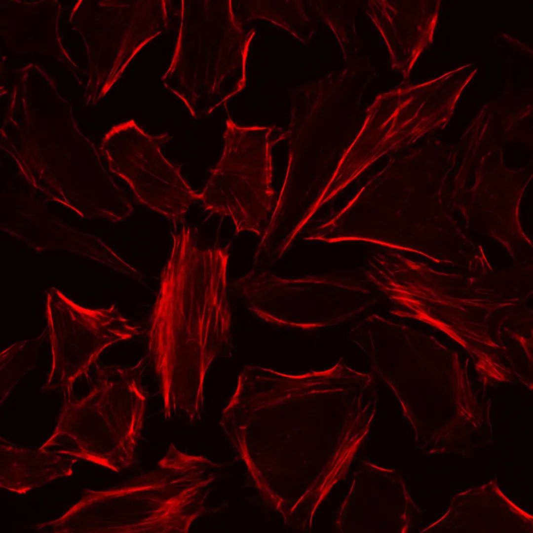
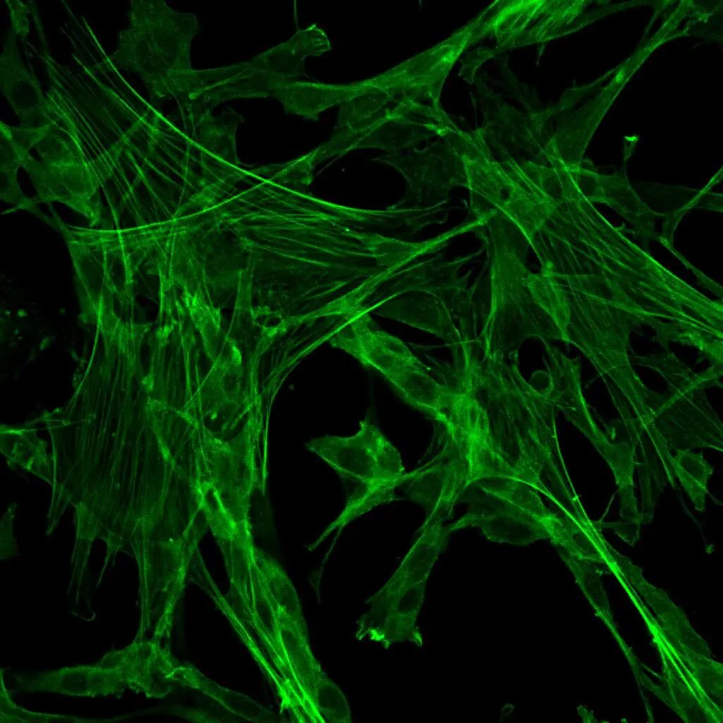
Lyso-Tracker Green (also known as DND-26) is a lysosome green fluorescent probe commonly used for specific staining of lysosomes in live cells. This probe consists of a fluorescent group linked to a weak base, allowing it to freely penetrate cells and accumulate within lysosomes, which have a weakly acidic interior. This results in effective labeling of lysosomes.
Storage and Transport:
Shipped on dry ice; store at -20°C, protect from light. The probe is stable for at least 12 months when stored properly.
Kit Components:
- Lyso-Tracker Green (Lysosome Green Fluorescent Probe) – 50 μL
- Product Manual – 1 copy
Operating Instructions:
- Preparation of Detection Solution:
1.1. The provided product is a 1 mM stock solution. Before use, warm it to room temperature, briefly centrifuge it at low speed to ensure it settles at the bottom of the tube.
1.2. Dilute the stock solution with buffer (such as Hanks’ balanced salt solution, PBS, or serum-free basal medium) to create a lysosome detection solution at a concentration of 50-100 nM. Adjust the concentration as needed, but use a lower concentration to avoid false positives due to high probe concentrations.
- Cell Staining (protocol for adherent cells, for suspension cells centrifugation is needed):
2.1. Wash normal or treated cells with buffer 1-2 times for 3-5 minutes each.
2.2. Add pre-warmed lysosome detection solution to cells in the cell culture incubator at 37°C for 30-60 minutes. Observe the fluorescence effect under a fluorescence microscope. Adjust the incubation time if necessary.
2.3. Remove the incubation solution and wash cells with buffer 2-3 times for 3-5 minutes each.
2.4. Observe the stained cells under a fluorescence microscope.
Precautions:
- Cell types and conditions may require adjustments in dye concentration and incubation time. Probe internalization dynamics suggest that probe entry into live cells takes only a few seconds. Note that the probe may cause lysosomal alkalization, resulting in an increase in lysosomal pH with prolonged incubation.
- This dye is suitable for lysosome localization in live cells and should not be used for subsequent fixation.
- Warm the detection solution and washing buffer to 37°C when staining and washing live cells. Staining and washing should be carried out in the cell culture incubator to avoid temperature changes that could affect cell morphology.
- The probe fluorescence is prone to photobleaching. Capture images promptly after staining and avoid exposure to light during the staining process.
- Store the probe stock solution in aliquots to prevent repeated freeze-thaw cycles. Prepare the staining solution as needed, using it immediately.
- Follow proper safety procedures, including wearing lab attire and gloves.
Note: The provided information is based on general knowledge about Lyso-Tracker Green and its typical usage. Refer to the specific product manual and instructions provided by the manufacturer for detailed and accurate protocols tailored to the product you are using.
This product is for research use only and is not suitable for clinical diagnosis.
Product Information
Product Name: Lyso-Tracker Green (Lysosome Green Fluorescent Probe) Product Number: Not provided in the text Specifications: Not provided in the text
Product Description:
Lyso-Tracker Green is a fluorescent dye commonly referred to as Lysosome Green Fluorescent Probe. It is used to specifically label lysosomes within cells. Lysosomes are membrane-bound organelles containing enzymes that break down waste materials and cellular components. Lyso-Tracker Green is designed to selectively stain lysosomes, enabling researchers to visualize and study their morphology, function, and dynamics.
Upon entering lysosomes, the probe accumulates and fluoresces, emitting green fluorescence. This allows for the observation of lysosomal structures using fluorescence microscopy setups and filters. Lyso-Tracker Green is widely used in cell biology research to investigate lysosomal processes and interactions within cells.
Storage and Transport:
Store at -20°C, protect from light. The probe should be stable for at least 6 months when stored properly.
Kit Components:
- Lyso-Tracker Green (provided amount not mentioned)
- Cell Staining Buffer (provided amount not mentioned)
- User Manual (1 copy)
Operating Instructions:
- Prepare Lyso-Tracker Green Staining Solution:
1.1. Dilute the Lyso-Tracker Green probe with Cell Staining Buffer to create the Lyso-Tracker Green Staining Solution. Adjust the dilution ratio based on specific conditions to achieve optimal staining.
- Staining Cells:
2.1. Prepare cells and place them in a suitable culture vessel.
2.2. Add the Lyso-Tracker Green Staining Solution to the culture medium at a concentration suitable for the experiment.
2.3. Incubate cells at 37°C, protected from light, for a recommended duration. Adjust the staining time based on experimental needs.
2.4. Wash cells with pre-warmed PBS to remove excess dye.
2.5. Observe stained cells under a fluorescence microscope. Lyso-Tracker Green emits green fluorescence.
Precautions:
- Fluorescent dyes may suffer from photobleaching. Minimize exposure to light to prolong fluorescence.
- Follow proper safety procedures, including wearing lab attire and gloves.
- Adjust the staining protocol based on cell type, experimental conditions, and specific research goals.
- The probe is intended for research use only and is not suitable for clinical diagnosis.
Note: The product information provided here is based on general knowledge about Lyso-Tracker Green and its typical usage. Please refer to the specific product manual and instructions provided by the manufacturer for detailed and accurate protocols tailored to the